. 2003 Aug 15;31(16):4917-28.
doi: 10.1093/nar/gkg671.

Cloning, functional analysis and post-transcriptional regulation of a type II DNA topoisomerase from Leishmania infantum. A new potential target for anti-parasite drugs

Affiliations

Cloning, functional analysis and post-transcriptional regulation of a type II DNA topoisomerase from Leishmania infantum. A new potential target for anti-parasite drugs

Tobias Hanke et al. Nucleic Acids Res. .

Abstract

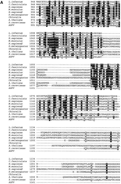
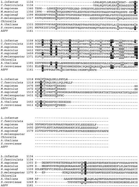
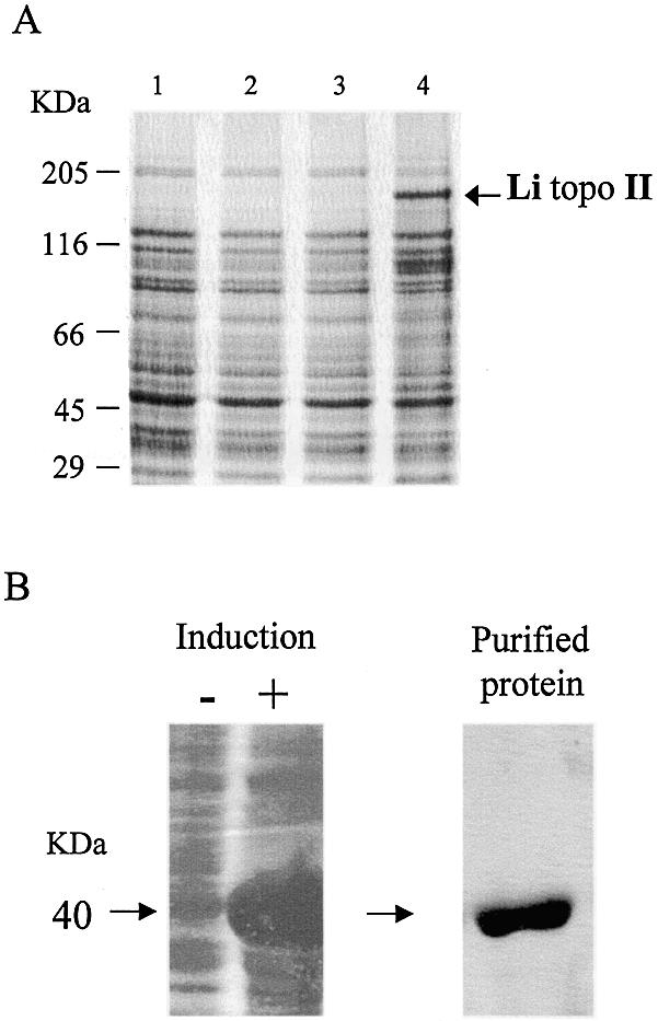
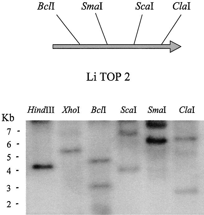
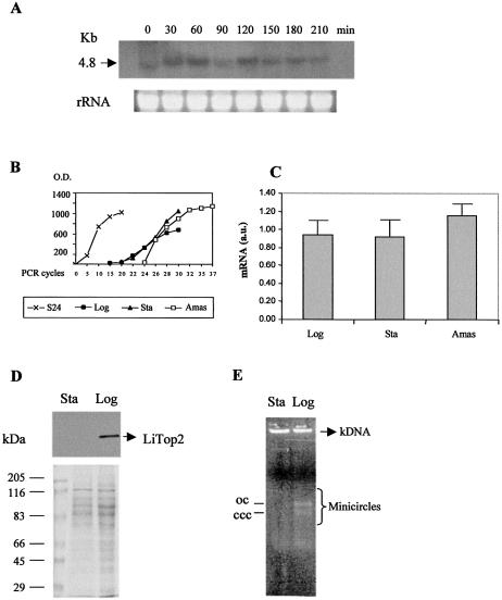
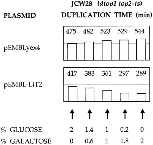
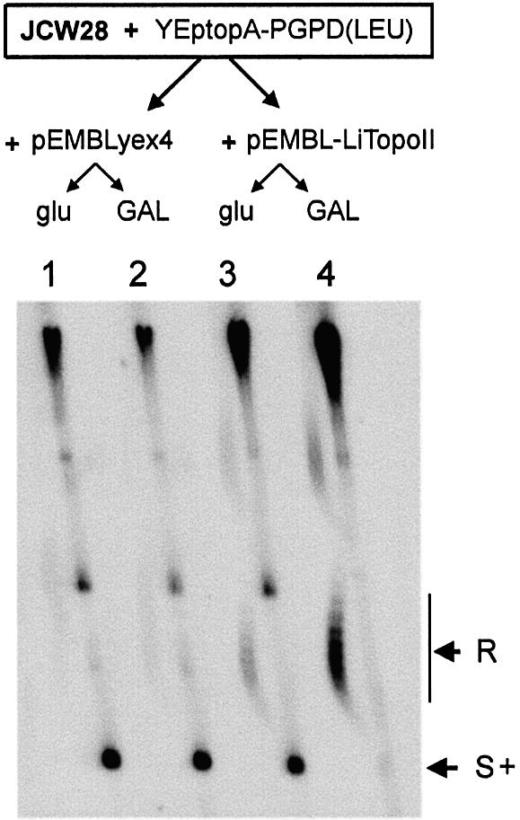
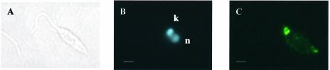
We identified a type II topoisomerase enzyme from Leishmania infantum, a parasite protozoon causing disease in humans. This protein, named Li topo II, which displays a variable C-terminal end, is located in the kinetoplast. The cloned gene encoding Li-TOP2 compensates for the slow growth of topo II-deficient mutants of Saccharomyces cerevisiae, resulting in a catalytically active DNA topoisomerase in yeast. Analysis of the specific mRNA levels of the Li-TOP2 gene showed variations throughout the parasite cell cycle in synchronized cells as well as between the distinct forms of the parasite. Thus, the enzyme had higher levels of mRNA expression in the highly infective intracellular form of the parasite, the amastigote, than in the extracellular promastigote form, suggesting a relation with the distinct developmental and infectious phases of the protozoon. In addition, western blot analysis showed differences in protein expression between the proliferative and non-proliferative forms of L.infantum promastigotes, which displayed similar levels of mRNA. This indicated possible post-transcriptional regulation mechanisms. The data suggest that Li topo II has a part in DNA decatenation and probably at the initial stages of proliferation in the intracellular form of L.infantum, a parasite that has to proliferate into the host macrophage to survive its hostile environment in its first moments of intracellular infection.


Figures

Figure 1
Figure 1
(A) Multiple amino acid alignment of C-terminal ends of topo II enzymes from different species. Black columns represent identical residues and gray columns represent homologous residues. (B) Phylogenetic trees showing the evolutionary distances between members of the topo II family. The species employed were: H.sapiens α isoform (NP001058); H.sapiens β isoform (NP_001059); M.musculus (NP035753); D.melanogaster (P15348); S.cerevisiae (P06786); L.infantum (AF86355); C.fasciculata (P27570); Chlorella (NP048939); and A.thaliana (NP189031). The multiple alignments were carried out using the CLUSTALW program. The phyletic trees derived from the multiple alignments were viewed by the Tree View program developed by Rod Page. For further experimental details, see Materials and Methods.
Figure 1
Figure 1
(A) Multiple amino acid alignment of C-terminal ends of topo II enzymes from different species. Black columns represent identical residues and gray columns represent homologous residues. (B) Phylogenetic trees showing the evolutionary distances between members of the topo II family. The species employed were: H.sapiens α isoform (NP001058); H.sapiens β isoform (NP_001059); M.musculus (NP035753); D.melanogaster (P15348); S.cerevisiae (P06786); L.infantum (AF86355); C.fasciculata (P27570); Chlorella (NP048939); and A.thaliana (NP189031). The multiple alignments were carried out using the CLUSTALW program. The phyletic trees derived from the multiple alignments were viewed by the Tree View program developed by Rod Page. For further experimental details, see Materials and Methods.
Figure 2
Figure 2
Li topo II over-expression and Li topo II-1.1 purification. (A) Coomassie blue staining of Li topo II over-expression. Lane 1, E.coli M15 strain; lane 2, E.coli M 15 strain harbored with plasmid pQE30; lane 3, E.coli M15 strain harbored with the plasmid pQE30-Li-topo II without IPTG; lane 4, E.coli M15 strain harbored with the plasmid pQE30-Li-topo II after 4 h of addition of IPTG. (B) Expression of Li topo II-1.1 polypeptide fragment (40 kDa) by an E.coli M15 strain harbored with the pQE30-Li topo II-1.1 plasmid before (not induced) and after (induced) the addition of IPTG and protein purification on Ni++-affinity resin. For further experimental details, see Materials and Methods.
Figure 3
Figure 3
Molecular gene analysis of Li-TOP2 gene. Southern blot analysis and physical map of Li-TOP2 gene deduced from its nucleotide sequence with the location of the restriction enzymes used. Leishmania infantum genomic DNA was subjected to total digestion with the indicated restriction enzymes. For further experimental details, see Materials and Methods.
Figure 4
Figure 4
Differential expression of Li topo II throughout the cell and parasite life cycle. (A) Northern blot analysis using 5 µg of total RNA extracted from L.infantum promastigotes taken from hydroxyurea-synchronous cell culture at different times (indicated in minutes). (B) Levels of expression by RT–PCR of S24α L.infantum ribosomal protein mRNA (control) and Li topo II mRNA from logarithmic (Log) and stationary (Sta) promastigotes as well as in intracellular amastigotes (Amas). (C) Relative levels of expression of Li topo II mRNA transcripts in the different L.infantum phases: logarithmic (Log), stationary (Sta) promastigotes and amastigotes (Amas). (D) Western blot analysis of the levels of Li topo II at the logarithmic and stationary phases of the parasite. (E) Decatenation activity assay of the Li topo II enzyme in cell extracts from the logarithmic (Log) and stationary (Sta) phases of L.infantum promastigotes. The positions of open circular (OC) and covalently closed circular (CCC) DNA products are indicated. For further experimental details, see Materials and Methods.
Figure 5
Figure 5
Growth rate of Δtop1 top2-4 yeast cells expressing Li topo II. Transformants of JCW28 cells, transformed either with pEMBLyex4 or pEMBL-LiTopoII, were grown in selective media SD (–ura) containing different amounts (w/v) of galactose and glucose as indicated. Duplication times (min) of each culture were measured during exponential growth.
Figure 6
Figure 6
DNA relaxation activity of L.infantum topoisomerase II in Δtop1 top2-4 yeast cells. Cultures containing 2% glucose or 2% galactose of JCW28 cells harboring YeptopA-PGPD(LEU) plus pEMBLyex4 (lanes 1 and 2) or pEMBL-LiTopoII (lanes 3 and 4) were set as described in the text. Cultures were shifted to 35°C for 2 h 30 min before cell harvesting. DNA extracts were examined by two-dimensional electrophoresis, carried out in a 0.6% agarose gel, that was run in TBE buffer plus 0.6 and 3 µg/ml chloroquine in the first (top to bottom, 12 h at 60 V) and second (left to right, 6 h at 60 V) dimensions, respectively. The gel was blot-hybridized with a 32P-labeled probe to reveal the 2µ yeast plasmid (6.3 kb). The positions of the arched distribution of topoisomerse and of the positively supercoiled forms (S+) of the 2µ are indicated. Bands appearing on the upper half of the gel mostly correspond to multimeric forms of the 2µ plasmid, chromosomal DNA fragments and less abundant DNA plasmids.
Figure 7
Figure 7
Kinetoplast localization of Li topo II by immunofluorescence. Leishmania infantum promastigotes were recovered from asynchronous cultures at the logarithmic phase. (A) Phase contrast. (B) DNA staining with DAPI shows the nucleus (n) and kinetoplast (k) localization at the promastigotes. (C) Li topo II at logarithmic phase promastigotes. Negative immunofluorescence in stationary phase promastigotes is not shown. Bar 2 µm. For further experimental details, see Materials and Methods.

Similar articles

Cited by

References

    1. Neva F. and Sacks,S. (1995) Leishmaniasis. In Warren,K.S. and Mahmoud,A.A.S. (eds), Leishmaniasis in Tropical and Geographical Medicine. McGraw-Hill, New York, pp. 296–308.
    1. Jimenez M.I., Gutierrez-Solar,B., Benito,A., Aguiar,A., García,E., Carcenado,E. and Alvar,J. (1991) Cutaneous Leishmania infantum zymodems isolated from bone marrow in AIDS patients. Res. Rev. Parasitol., 51, 91–95.
    1. Sacks D.L. and Perkins,P.V. (1984) Identification of an infective stage of Leishmania promastigotes. Science, 223, 1417–1419. - PubMed
    1. Da Silva R. and Sacks,D.L. (1987) Metacyclogenesis is a major determinant of Leishmania promastigote virulence and attenuation. Infect. Immun., 55, 2802–2806. - PMC - PubMed
    1. Soguin M.L., Elwood,H.J. and Gunderson,J.H. (1986) Evolutionary diversity of eukaryotic small-subunit rRNA genes. Proc. Natl Acad. Sci. USA, 83, 1383–1387. - PMC - PubMed

Publication types

MeSH terms