Comparative Study
. 2003 Aug 15;31(16):4950-7.
doi: 10.1093/nar/gkg672.

Characterisation of the substrate specificity of homogeneous vaccinia virus uracil-DNA glycosylase

Affiliations
Comparative Study

Characterisation of the substrate specificity of homogeneous vaccinia virus uracil-DNA glycosylase

Natale Scaramozzino et al. Nucleic Acids Res. .

Abstract

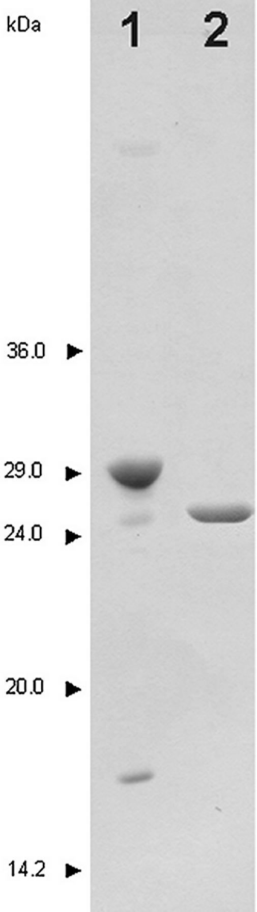
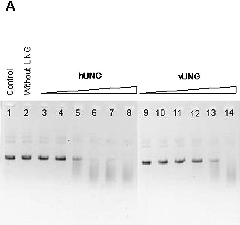
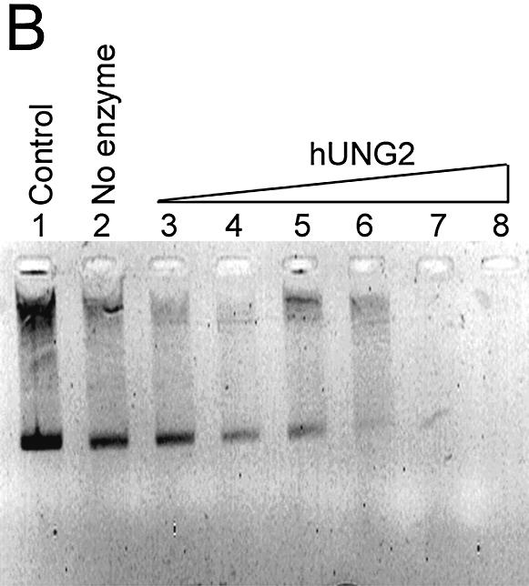
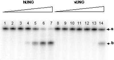
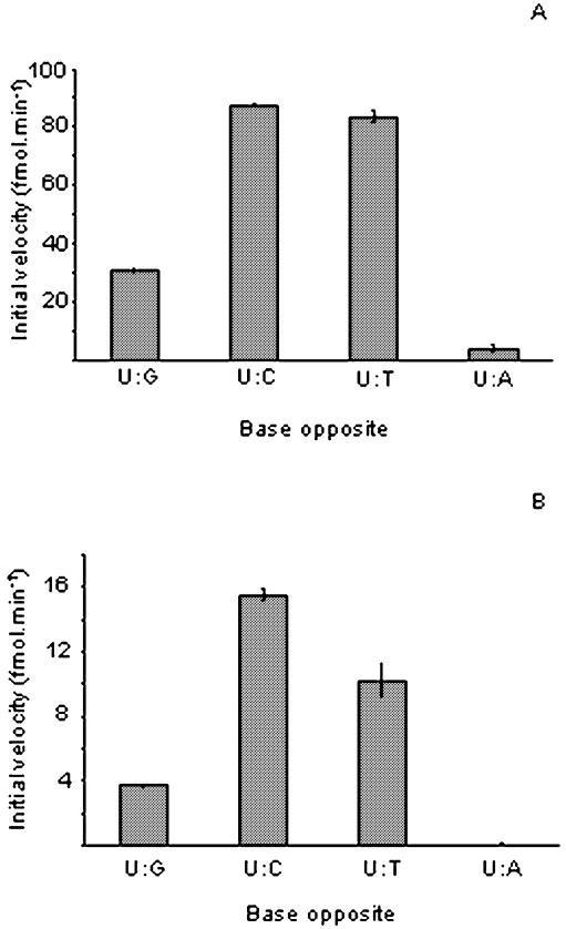
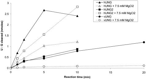
The decision to stop smallpox vaccination and the loss of specific immunity in a large proportion of the population could jeopardise world health due to the possibility of a natural or provoked re-emergence of smallpox. Therefore, it is mandatory to improve the current capability to prevent or treat such infections. The DNA repair protein uracil-DNA glycosylase (UNG) is one of the viral enzymes important for poxvirus pathogenesis. Consequently, the inhibition of UNG could be a rational strategy for the treatment of infections with poxviruses. In order to develop inhibitor assays for UNG, as a first step, we have characterised the recombinant vaccinia virus UNG (vUNG) and compared it with the human nuclear form (hUNG2) and catalytic fragment (hUNG) UNG. In contrast to hUNG2, vUNG is strongly inhibited in the presence of 7.5 mM MgCl(2). We have shown that highly purified vUNG is not inhibited by a specific uracil-DNA glycosylase inhibitor. Interestingly, both viral and human enzymes preferentially excise uracil when it is opposite to cytosine. The present study provides the basis for the design of specific inhibitors for vUNG.


Figures

Figure 1
Figure 1
SDS–PAGE analysis of the purified recombinant hUNG and vUNG. Details of the purification are described in Materials and Methods. Lane 1, vUNG (1 µg); lane 2, hUNG (1 µg). The molecular weight standards were rabbit muscle glyceraldehyde-3-phosphate dehydrogenase (36 kDa), bovine erythrocytes carbonic anhydrase (29 kDa), bovine pancreas trypsinogen (24 kDa), soybean trypsin inhibitor (20 kDa) and bovine milk α-lactalbumin (14.2 kDa). Proteins were stained with Coomassie blue R250.
Figure 2
Figure 2
Activity of vUNG, hUNG and hUNG2 on single-stranded plasmid DNA containing uracil residues [pBluescript II KS(–)]. pBluescript II KS(–) (0.25 µg) was incubated with various concentrations of vUNG, hUNG and hUNG2 as described in Materials and Methods. Reaction products were separated by electrophoresis in a 0.8% agarose gel and stained with ethidium bromide. (A) Lane 1, pBluescript II KS(–) control; lane 2, as lane 1 but with 20 nM Fpg; lanes 3–8, as lane 2 but treated with increasing amounts of hUNG; lanes 9–14, as lane 2 but treated with increasing amounts of vUNG. The UNG protein concentrations were 0.001 nM in lanes 3 and 9; 0.01 nM in lanes 4 and 10; 0.1 nM in lanes 5 and 11; 1 nM in lanes 6 and 12; 10 nM in lanes 7 and 13; 100 nM in lanes 7 and 14. (B) Lane 1, pBluescript II KS(–) control; lane 2, as lane 1 but treated with piperidine; lanes 3–8, as lane 2 but treated with increasing amounts of hUNG2; lane 3, 0.001 nM; lane 4, 0.01 nM; lane 5, 0.1 nM; lane 6, 1 nM; lane 7, 10 nM; lane 8, 100 nM.
Figure 2
Figure 2
Activity of vUNG, hUNG and hUNG2 on single-stranded plasmid DNA containing uracil residues [pBluescript II KS(–)]. pBluescript II KS(–) (0.25 µg) was incubated with various concentrations of vUNG, hUNG and hUNG2 as described in Materials and Methods. Reaction products were separated by electrophoresis in a 0.8% agarose gel and stained with ethidium bromide. (A) Lane 1, pBluescript II KS(–) control; lane 2, as lane 1 but with 20 nM Fpg; lanes 3–8, as lane 2 but treated with increasing amounts of hUNG; lanes 9–14, as lane 2 but treated with increasing amounts of vUNG. The UNG protein concentrations were 0.001 nM in lanes 3 and 9; 0.01 nM in lanes 4 and 10; 0.1 nM in lanes 5 and 11; 1 nM in lanes 6 and 12; 10 nM in lanes 7 and 13; 100 nM in lanes 7 and 14. (B) Lane 1, pBluescript II KS(–) control; lane 2, as lane 1 but treated with piperidine; lanes 3–8, as lane 2 but treated with increasing amounts of hUNG2; lane 3, 0.001 nM; lane 4, 0.01 nM; lane 5, 0.1 nM; lane 6, 1 nM; lane 7, 10 nM; lane 8, 100 nM.
Figure 3
Figure 3
Activity of vUNG and hUNG on single-stranded oligonucleotide containing a single uracil residue (PS-U). 5′ 32P-labelled PS-U (10 nM) was incubated with various amounts of UNG at 37°C for 5 min. Reaction products were subjected to light piperidine treatment to reveal abasic sites generated by DNA glycosylase action. They were then separated by electrophoresis on a 20% polyacrylamide gel containing 7 M urea and visualised using a PhosphorImager Storm 840. Lanes 1–7, PS-U treated with increasing amounts of hUNG; lanes 8–14, PS-U treated with increasing amounts of vUNG. Lanes 1 and 8, PS-U control. UNG protein concentrations were 0.001 nM in lanes 2 and 9; 0.01 nM in lanes 3 and 10; 0.1 nM in lanes 4 and 11; 1 nM in lanes 5 and 12; 10 nM in lanes 6 and 13; and 100 nM in lanes 7 and 14. a, 45mer PS-U; b, 20mer cleaved products.
Figure 4
Figure 4
Cleavage of duplex oligonucleotides containing different bases opposite to uracil by hUNG and vUNG. The 5′ 32P-labelled PS-U oligonucleotide was annealed to various complements to generate the following mismatches: U·G, U·C, U·T and U·A. Each duplex oligonucleotide (10 nM) was incubated with hUNG (1 nM) (A) and vUNG (50 nM) (B) at 37°C for various periods of time (1–20 min). Reaction products were subjected to light piperidine treatment to reveal abasic sites generated by UNG and analysed as described in Materials and Methods.
Figure 5
Figure 5
Effect of MgCl2 on the activity of the vUNG, hUNG and hUNG2 proteins. The U·G duplex oligonucleotide (250 nM) was incubated with hUNG (1 nM) and vUNG (50 nM) at 37°C for 1–20 min in the presence or absence of 7.5 mM MgCl2. Reaction products were subjected to light piperidine treatment to reveal abasic sites generated by UNG and analysed as described in Materials and Methods. The radioactivity of the reaction products was quantified and plotted against time.

Similar articles

Cited by

References

    1. Goebel S.J., Johnson,G.P., Perkus,M.E., Davis,S.W., Winslow,J.P. and Paoletti,E. (1990) The complete DNA sequence of vaccinia virus. Virology, 179, 247–266, 517–263. - PubMed
    1. Moss B. (2001) Poxviridae: the virus and their replication. In Knipe,D.M. and Howley,P.M. (eds), Fields Virology. Raven Press, New York, Vol. 2, pp. 2849–2884.
    1. Traktman P. (1990) Poxviruses: an emerging portrait of biological strategy. Cell, 62, 621–626. - PubMed
    1. Moss B., Ahn,B.Y., Amegadzie,B., Gershon,P.D. and Keck,J.G. (1991) Cytoplasmic transcription system encoded by vaccinia virus. J. Biol. Chem., 266, 1355–1358. - PubMed
    1. Traktman P. (1996) Poxvirus DNA replication. In DePamphilis,M.L. (ed.), DNA Replication in Eucaryotic Cells. Cold Spring Harbor Laboratory Press, New York, pp. 775–793.

Publication types

MeSH terms