Comparative Study
. 2003 Aug 15;31(16):e88.
doi: 10.1093/nar/gng088.

Dendrimeric coating of glass slides for sensitive DNA microarrays analysis

Affiliations
Comparative Study

Dendrimeric coating of glass slides for sensitive DNA microarrays analysis

Véronique Le Berre et al. Nucleic Acids Res. .

Abstract

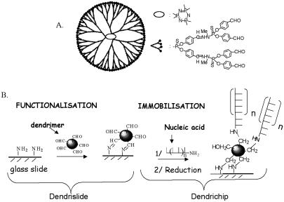
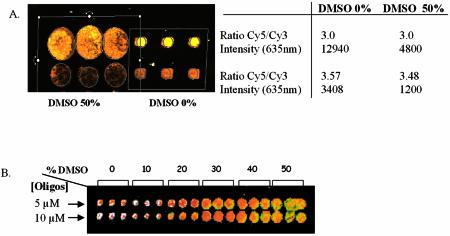
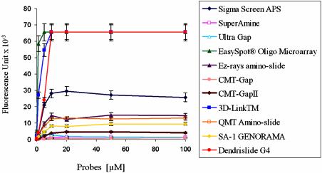
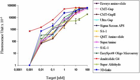
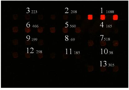
Successful use and reliability of microarray technology is highly dependent on several factors, including surface chemistry parameters and accessibility of cDNA targets to the DNA probes fixed onto the surface. Here, we show that functionalisation of glass slides with homemade dendrimers allow production of more sensitive and reliable DNA microarrays. The dendrimers are nanometric structures of size-controlled diameter with aldehyde function at their periphery. Covalent attachment of these spherical reactive chemical structures on amino-silanised glass slides generates a reactive approximately 100 A layer onto which amino-modified DNA probes are covalently bound. This new grafting chemistry leads to the formation of uniform and homogenous spots. More over, probe concentration before spotting could be reduced from 0.2 to 0.02 mg/ml with PCR products and from 20 to 5 micro M with 70mer oligonucleotides without affecting signal intensities after hybridisation with Cy3- and Cy5-labelled targets. More interestingly, while the binding capacity of captured probes on dendrimer-activated glass surface (named dendrislides) is roughly similar to other functionalised glass slides from commercial sources, detection sensitivity was 2-fold higher than with other available DNA microarrays. This detection limit was estimated to 0.1 pM of cDNA targets. Altogether, these features make dendrimer-activated slides ideal for manufacturing cost-effective DNA arrays applicable for gene expression and detection of mutations.


Figures

Figure 1
Figure 1
Scheme of dendrimer ‘generation 4’ (A) and description of surface activation to fix the dendrimers and immobilisation method for covalent binding of nucleic acids (DNA probes) on the activated glass slide surface (B).
Figure 2
Figure 2
Images of hybridisation of a DNA probe spotted on activated glass slides with dendrimer generation 4 in the absence or in the presence of DMSO. (A) PCR products from two yeast ORFs (CDC19/YAL038w and CLN3/YAL040c) were resuspended in Na2 phosphate buffer 0.3 M, pH 9 in the absence or in the presence of DMS0 50%. (B) A 5′-NH2 35mer oligonucleotide was prepared in the same buffer at two different concentrations (5 and 10 µM) in the absence or presence of increasing concentration of DMSO. DNAs were spotted in triplicate on the activated dendrislides and hybridisation was carried out with yeast labelled cDNA (A) or with a complementary Cy5-labelled 15mer (5′-Cy5-AATGGTATCGGAGTA-3′) (B).
Figure 3
Figure 3
Image capture from hybridisation of arrays manufactured with dendrislide bearing DNA probes spotted at decreasing concentrations. (A) PCR from yeast ORF (ORF1: MET14/YKL001c, ORF2: SPC42/YKL042w, ORF3: MRP17/YKL003c) was spotted in duplicate (column) in phosphate buffer 0.3 M, pH 9 at 200 and 20 µg/ml (rows) in the absence or in the presence of 1.5 M betaine. (B) A 70mer oligonucleotide corresponding to the gene SSA1/YAL005c was spotted in quadruplet in the same buffer at decreasing concentration ranging from 20 to 1 µM. The hybridisation was carried out with Cy3-labelled cDNA (A) or a mix of Cy3-/Cy5-labelled cDNA (B) obtained from 10 µg of yeast total RNA. In (A), the value of fluorescence intensity (fluorescent signal – background) measured at 532 nm is given on the top of the corresponding spots. The fluorescence intensity was measured with the Genepix scanner at 500 PMT.
Figure 4
Figure 4
Immobilisation efficiency of G4-dendrislides compared with other commercially available activated-glass slides. Single-stranded 35mer oligonucleotide carrying a 3′-amino function and a 5′-Cy5 fluorophore (5′-Cy5-TTAGCGCATTTTGGCATATTTGG GCGGACAACTT-NH2-3′) was spotted at concentrations from 0.01 to 100 µM, on the different functionalised-glass slides. The value of fluorescence intensity (fluorescent signal – background) was measured at 635 nm with the Genepix scanner at 500 PMT.
Figure 5
Figure 5
Detection sensitivity of dendrislide compared with commercially available activated glass slides. A single-stranded 35mer amino-oligonucleotide (5′-NH2-GTGATCGTTGTATCGAGGAATACTCCGATACCATT-3′) at a concentration of 10 µM was spotted on the different functionalised glass slides. The target Cy5-labelled 15mer oligonucleotides (5′-Cy5-AATGGTATCGGAGTA-3′) were hybridised at concentrations ranging from 0.001 to 100 nM. Image capture was measured at 635 nm with GenePix 4000A at 500 PMT. The value of fluorescence intensity reported is the fluorescent signal – background.
Figure 6
Figure 6
Use of dendrislides to detect base mutations. Thirteen 20mer oligonucleotides were spotted in triplicate at 10 µM in Pi buffer 0.3 M, pH 9.0. These oligonucleotides corresponded to a part of the yeast HSP12 gene, and varied from each other by a single or two mutations proximal to the 5′- or 3′-end or in the middle of the sequence. The 20mer sequences from HSP12 were as follows: 1: NH2 5′-AATATGTTTCCGGTCGTGTC-3′; 2: NH2 5′-AATATGTTTCGGGTCGTGTC-3′; 3: NH2 5′-AATATGTT TCAGGTCGTGTC-3′; 4: NH2 5′-AATATGTTTCtGGTCGTGTC-3′; 5: NH2 5′-AATATGTTTCCGGTCGTGTA-3′; 6: NH2 5′-AATATGTTTCC GGTCGTGTG-3′; 7: NH2 5′-AATATGTTTCCGGTCGTGTT-3′; 8: NH2 5′-AATATGATTCCGGaCGTGTC-3′; 9: NH2 5′-AATATGGTTCCGG GCGTGTC-3′; 10: NH2 5′-AATATGCTTCCGGcCGTGTC-3′; 11: NH2 5′-AATAAGTTTCCGGTCGTGTC-3′; 12: NH2 5′-AATAGGTTTCCGGT CGTGTC-3′; 13: NH2 5′-AATACGTTTCCGGTCGTGTC-3′. Hybridisation was carried out with Cy5-labelled cDNA from yeast total RNA. At the top of the corresponding spots is given the mean value of fluorescent signal (intensity at 635 nm – background).

References

    1. Lipshutz R.J., Fodor,S.P.A., Gingeras,T.R. and Lockhart,D.J. (1999) High density synthetic oligonucleotide arrays. Nature Genet., 14, 1675–1680. - PubMed
    1. Hughes T.R., Mao,L.M., Jones,A.R., Burchard,J., Marton,M.J., Shannon,K.W. et al. (2001) Expression profiling using microarrays fabricated by an ink-jet oligonucleotide synthesizer. Nat. Biotechnol., 19, 342–347. - PubMed
    1. Cheung V.G., Morley,M., Auilar,F., Massimi,A., Kucherlapati,R. and Childs,G. (1999) Making and reading microarrays. Nature Genet., 21, (suppl.), 15–19. - PubMed
    1. MacBeath G., Koehler,A.N. and Schreiber,S.L.(1999) Printing small molecules as microarrays and detecting protein-ligand interactions en masse. J. Am. Chem. Soc., 121, 7967–7968.
    1. Mac Beath G. and Schreiber,S.L. (2000) Printing proteins as microarrays for high-throughput function determination. Science, 289, 1760–1762. - PubMed

Publication types

MeSH terms