Comparative Study
. 2003 Aug 15;31(16):e90.
doi: 10.1093/nar/gng090.

Combination of DNA-directed immobilization and immuno-PCR: very sensitive antigen detection by means of self-assembled DNA-protein conjugates

Affiliations
Comparative Study

Combination of DNA-directed immobilization and immuno-PCR: very sensitive antigen detection by means of self-assembled DNA-protein conjugates

Christof M Niemeyer et al. Nucleic Acids Res. .

Abstract

An assay for very sensitive antigen detection is described which takes advantage of the self- assembly capabilities of semi-synthetic conjugates of DNA and proteins. The general scheme of this assay is similar to a two-sided (sandwich) enzyme-linked immunoassay (ELISA); however, covalent single-stranded DNA-streptavidin (STV) conjugates, capable of hybridizing to complementary surface-bound DNA oligomers, are utilized for the effective immobilization of either capture antibodies or antigens, rather than the chemi- or physisorption usually applied in ELISA. Immuno-PCR (IPCR) is employed as a method for signal generation, utilizing oligomeric reagents obtained by self-assembly of STV, biotinylated DNA and antibodies. In three different model systems, detecting human IgG, rabbit IgG or carcinoembryonic antigen, this combination allowed one to increase the sensitivity of the analogous ELISA approximately 1000-fold. For example, <0.1 amol/ micro l (15 pg/ml) of rabbit IgG was detectable. The immunoassay can be carried out in a single step by tagging the analyte with both reagents for capture and read-out simultaneously, thereby significantly reducing handling time and costs of analysis. Moreover, as the spatial selectivity of target immobilization is determined by the specificity of DNA base pairing, the assay is particularly suited for miniaturized microfluidics and lab-on-a-chip devices.


Figures
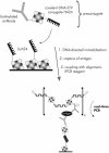
Figure 1
Figure 1
Schematic representation of the immunoassay based on DDI and IPCR. A covalent DNA–STV conjugate, HA24, is coupled with a biotinylated antibody by mixing the two compounds, thereby generating a preconjugate. Biotinylated antisense capture-oligonucleotide bcA24 was immobilized on STV-coated microplates and the remaining free biotin-binding sites of the surface-bound STV are blocked with D-biotin, represented by shaded spheres. The preconjugates are then allowed to bind to their complement by specific DNA hybridization. The immobilized antibody is used for capturing the antigen and the amount of target analyte is determined by IPCR, using oligomeric conjugates comprised of STV, bis-biotinylated dsDNA and biotinylated antibodies. The read-out of immunoassay is carried out using real-time PCR.
Figure 2
Figure 2
Real-time PCR amplification curves of the DDI–IPCR assay for the detection of rabbit IgG (rIgG). Representative samples of different concentrations of the rIgG antigen, analyzed in duplicate by the DDI–IPCR assay, are shown. The TaqMan software calculates the threshold cycle at which the fluorescent reporter signal exceeds the signal of the threshold. This threshold cycle is set at 100 ΔRn. Data points A–D represent different rIgG concentrations of 10 nM (A), 100 pM (B), 1 pM (C) and 10 fM (D), respectively. E indicates the NC of the IPCR (no rIgG antigen present during immuno-capture) and F the NC of the PCR (amplification of pure PCR reagent without the presence of any template). Note the high reproducibility of the real-time curves with a standard deviation of <8% (mean 1.6%) for duplicate measurements. Four-fold independent repetition of this assay, carried out under identical conditions, also revealed an inter-experimental error of <8% (mean 4.5%).
Figure 3
Figure 3
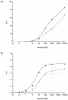
DDI of capture antibodies from goat directed against human IgG (GAH). Immobilization efficiencies of three alternative techniques are compared: biotinylated GAH was immobilized by either direct physisorption (triangles), biotin–STV interaction (rectangles) or DDI (circles). Serial dilutions of the target antigen (human IgG, hIgG) were incubated in the wells containing the capture antibody. Signal detection was carried out by either regular ELISA (A), using an anti-hIgG–STV–alkaline phosphatase conjugate and fluorescence detection or IPCR (B) using an anti-human IgG-STV-DNA conjugate and real-time TaqMan detection. The standard deviation of duplicate measurements was <5%, e.g. 1.04 ± 0.016 for detection of 1 amol/µl hIgG (B, closed circles). Note the ∼100-fold increase in sensitivity associated with the use of IPCR instead of ELISA signal detection.
Figure 4
Figure 4
Application of the DDI–IPCR combined assay for the detection of CEA. The signals obtained for serial dilutions of CEA in human blood serum are compared for two different assays, i.e. the DDI–ELISA (light gray) and DDI–IPCR (black). The standard deviation in IPCR is typically <5%, e.g. 1.16 ± 0.006 for the detection of 5 amol/µl CEA (black). Note that IPCR detection leads to an ∼1000-fold enhancement in the limit of detection.
Figure 5
Figure 5
Detection of IgG from human (hIgG, black rectangles) and rabbit (rIgG, light-gray circles) by means of the DDI–IPCR assay. The standard deviation of duplicate measurements was <8%, e.g. 1.31 ± 0.093 for detection of 1 amol/µl rIgG (gray circles). Note the increased sensitivity for the detection of rIgG, as compared with hIgG.
Figure 6
Figure 6
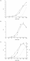
Performance of DDI-based in-solution capture assays for the detection of rIgG. (A) Two-step immunoassay: serial dilutions of the rIgG analyte were mixed with varying amounts of capture reagent HA24-GAR, ranging from 2 (rectangles), 10 (circles) to 50 nM (triangles). The immuno-complexes formed in solution were immobilized in DNA-functionalized microplates, IPCR detection conjugate was bound, and signal detection was achieved by real-time PCR. (B) One-step immunoassay, carried out by mixing serial dilutions of rIgG with a fixed amount of capture reagent (circles, 4 nM; rectangles, 10 nM) and IPCR detection conjugate (25 pM). (C) Comparison of the three different DDI–IPCR assays investigated, requiring either three steps (rectangles), two steps (triangles) or one step (circles) of reagent incubation. The two- and three-step assays were obtained using 10 nM of capture reagent, while 4 nM was used in the one-step assay. Note that the standard deviation is typically <8%, e.g. 1.25 ± 0.07 for the detection of 1 amol/µl rIgG in the one-step assay (C, circles).

References

    1. Khandurina J. and Guttman,A. (2002) Bioanalysis in microfluidic devices. J. Chromatogr. A, 943, 159–183. - PubMed
    1. Burke D.T., Burns,M.A. and Mastrangelo,C. (1997) Microfabrication technologies for integrated nucleic acid analysis. Genome Res., 7, 189–197. - PubMed
    1. Quake S.R. and Scherer,A. (2000) From micro- to nanofabrication with soft materials. Science, 290, 1536–1540. - PubMed
    1. Thorsen T., Maerkl,S.J. and Quake,S.R. (2002) Microfluidic large-scale integration. Science, 298, 580–584. - PubMed
    1. Pirrung M.C. (2002) How to make a DNA chip. Angew. Chem. Int. Ed., 41, 1276–1289. - PubMed

Publication types

MeSH terms