. 2003 Jul;20(7):681-95.
doi: 10.1089/089771503322144590.

Median nerve trauma in a rat model of work-related musculoskeletal disorder

Affiliations

Median nerve trauma in a rat model of work-related musculoskeletal disorder

Brian D Clark et al. J Neurotrauma. 2003 Jul.

Abstract

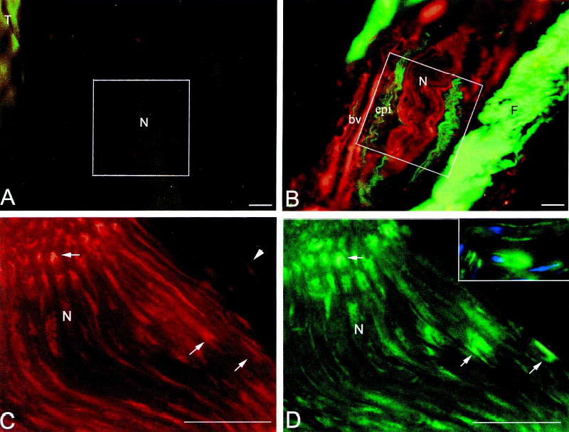
Anatomical and physiological changes were evaluated in the median nerves of rats trained to perform repetitive reaching. Motor degradation was evident after 4 weeks. ED1-immunoreactive macrophages were seen in the transcarpal region of the median nerve of both forelimbs by 5-6 weeks. Fibrosis, characterized by increased immunoexpression of collagen type I by 8 weeks and connective tissue growth factor by 12 weeks, was evident. The conduction velocity (NCV) within the carpal tunnel showed a modest but significant decline after 9-12 weeks. The lowest NCV values were found in animals that refused to participate in the task for the full time available. Thus, both anatomical and physiological signs of progressive tissue damage were present in this model. These results, together with other recent findings indicate that work-related carpal tunnel syndrome develops through mechanisms that include injury, inflammation, fibrosis and subsequent nerve compression.


Figures

FIG. 1
FIG. 1
Behavioral outcomes at weekly endpoints from the first week of the task regimen (week 1) through week 11, expressed as mean + SEM. (A) Observed reach rate versus week. There was a significant decrease in reach rate in weeks 5 and 6 compared to week 1 (p < 0.05). There was a rebound toward baseline (week 1) reach rate in week 8, followed by another decline in weeks 9–11 (not significant, possibly due to the decreasing numbers of subjects over time). (B) Task duration versus week. There was a significant decline in task duration in weeks 3 and 4 (p < 0.05), with a return toward baseline thereafter. Again, the lack of statistical significance in later weeks is possibly the result of decreasing numbers of subjects over time. *Significantly different from week 1 (p < 0.05). Numbers (n) of animals: week 1, n = 39; week 2, n = 38; week 3, n = 36; week 4, n = 33; week 5, n = 30; week 6, n = 27; week 7, n = 21; week 8, n = 19; week 9, n = 8; and weeks 10–11, n = 4.
FIG. 2
FIG. 2
Increase in ED1+ macrophages and degraded MBP in median nerves after 12 weeks of the task regimen. MBP and neurofilament 200-kD immunoreactivities are shown for comparison. (A) Only a few ED1+ cells (arrow) are visible in the nerve at the level of the carpal ligament at week 0. (B) Several ED1+ cells (arrows) are visible in the same region of the reach limb by 6 weeks. (C) In the same limb as B but more distal, many ED1+ cells (arrows) are present in nerve branches and surrounding connective tissue. (D) At week 12, scant degraded MBP immunoreactivity is visible in the nerve at the mid-forearm level. (E) Numerous whole MBP+ profiles are visible in the same section as (D). (F) Same section showing double staining for both anti-degraded MBP and anti-MBP. (G) At week 12, degraded MBP+ is greatly increased in the median nerve at the level of the wrist. The large cell (arrowhead) containing degraded MBP immunoreactivity may be a phagocytic macrophage. (H) Numerous whole MBP+ profiles are visible in the same section as G. The inset in H shows no overlap between MPB and neurofilament (blue) immunoreactivity. (I) Same section showing double staining for anti-degraded MBP and anti-MBP. There is only a small amount of staining overlap. N, nerve; CT, connective tissue. Bar = 50 μm.
FIG. 3
FIG. 3
Quantification of histological analyses, expressed as mean + SEM. (A) Quantification of the mean numbers of ED1+ macrophages in the median nerve at the level of the wrist and immediately proximal and distal to the carpal ligament. There are significantly more ED1+ macrophages in both the reach and nonreach limbs at 5/6, 8, and 12 weeks compared to week 0. The increases are greater in the reach than in the non-reach limbs. Numbers (n) of animals: week 0, n = 7; week 3/4, n = 7; week 5/6, n = 5; week 8, n = 4; and week 12, n = 4. (B) Quantification of area fraction containing collagen type I immunoreactivity in the same regions described in A. The area fraction of collagen type I immunoreactivity is significantly greater in both the reach and nonreach limbs at weeks 8 and 12 compared to week 0. Collagen type I immunoreactivity does not increase in the nonreach limbs until week 12; that increase is lower than in the reach limbs. Numbers of animals: week 0, n = 3; week 3/4, n = 4; week 5/6, n = 3; week 8, n = 3; and week 12, n = 4. (C) Quantification of area fraction containing CTGF immunoreactivity in the same regions described in A. The area fraction of CTGF immunoreactivity is significantly greater in both the reach and nonreach limbs at week 12 than at week 0. This increase is greater in the reach than in the nonreach limbs. Numbers of animals were the same as in B. **p < 0.001.
FIG. 4
FIG. 4
Macroscopic photographs of the median nerve traversing the carpal region of a week 0 (A) and a week 10 (B) rat. Note the connective tissue (CT) nodule surrounding the nerve in the reach limb of a rat that performed the task for 10 weeks.
FIG. 5
FIG. 5
Expression of collagen type I (A,B; green), and CTGF (A,B,C,D inset; red) increase in the median nerve at the level of the wrist with task performance. Immunofluorescence co-localization of CTGF and S100β (D,C inset; green) was used to detect cell-type specific expression of CTGF. (A) Little or no deposition of collagen type I is seen in the epineurium of the median nerve (N) at 0 weeks. (B) Increased collagen type I immunoreactivity is seen in the epineurium (epi) of the median nerve in the reach limb of a 12-week rat. F, fibrotic connective tissue with increased collagen type I staining. (C) CTGF is expressed by Schwann cells (representative profiles indicated by arrows) located within the nerve fascicles of a median nerve from the reach limb of 12-week rat. It may also be present in axons. Arrowheads indicate CTGF+ (S100β-; D) cells that may be epineurial fibroblasts. (D) The same section as C showing S100β expression by Schwann cells (representative profiles indicated by arrows) in the nerve fascicles. The thickness (16 μm) of these longitudinal sections captures both cross-sectional and surface profiles of Schwann cell cytoplasm. Inset shows no co-localization between S100β and neurofilament (blue) immunoreactivity. bv, blood vessel; epi, epineurium; F, fibrotic connective tissue; N, nerve, T, tendon. *The same cell in C, C inset, and D. Bar = 50 μm.
FIG. 6
FIG. 6
Plots of transcarpal median nerve conduction velocity. (A) Frequency histogram of NCV in median nerves of 30 control limbs (dotted outline) and the reach limbs of seven rats after 9–12 weeks of task performance (gray, solid outline). (B) Relationship between duration of task performance in the final week of the task regimen and NCV in the seven rats. The arrow indicates the duration of performance of the animal shown in Figure 4, for which the NCV could not be measured.

Similar articles

Cited by

References

    1. AVELLINO AM, HART D, DAILEY AT, et al. Differential macrophage responses in the peripheral and central nervous system during Wallerian degeneration of axons. Exp Neurol. 1995;136:183–198. - PubMed
    1. BACKMAN C, BOQUIST L, FRIDEN J, et al. Chronic achilles paratenonitis with tendinosis: an experimental model in the rabbit. J Orthop Res. 1990;8:541–547. - PubMed
    1. BARBE MB, BARR AE, GORZELANY I, et al. Chronic repetitive reaching and grasping results in decreased motor performance and widespread tissue responses. J Orthop Res. 2003;21:167–176. - PMC - PubMed
    1. BARR AE, SAFADI FF, GARVIN RP, et al. Evidence of progressive tissue pathophysiology and motor behavior degradation in a rat model of work related musculoskeletal disease. Proc Int Ergonomics Assoc Human Factors Ergonomics Soc. 2000;5:584–587.
    1. BARR AE, BARBE MF. Pathophysiologicaltissue changes associated with repetitive movement: a review of the evidence. Phys Ther. 2002;82:173–187. - PMC - PubMed

Publication types

LinkOut - more resources