Median nerve trauma in a rat model of work-related musculoskeletal disorder
- PMID: 12908929
- PMCID: PMC1550513
- DOI: 10.1089/089771503322144590
Median nerve trauma in a rat model of work-related musculoskeletal disorder
Abstract
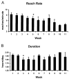
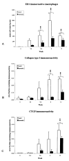
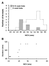
Anatomical and physiological changes were evaluated in the median nerves of rats trained to perform repetitive reaching. Motor degradation was evident after 4 weeks. ED1-immunoreactive macrophages were seen in the transcarpal region of the median nerve of both forelimbs by 5-6 weeks. Fibrosis, characterized by increased immunoexpression of collagen type I by 8 weeks and connective tissue growth factor by 12 weeks, was evident. The conduction velocity (NCV) within the carpal tunnel showed a modest but significant decline after 9-12 weeks. The lowest NCV values were found in animals that refused to participate in the task for the full time available. Thus, both anatomical and physiological signs of progressive tissue damage were present in this model. These results, together with other recent findings indicate that work-related carpal tunnel syndrome develops through mechanisms that include injury, inflammation, fibrosis and subsequent nerve compression.
Figures


References
-
- AVELLINO AM, HART D, DAILEY AT, et al. Differential macrophage responses in the peripheral and central nervous system during Wallerian degeneration of axons. Exp Neurol. 1995;136:183–198. - PubMed
-
- BACKMAN C, BOQUIST L, FRIDEN J, et al. Chronic achilles paratenonitis with tendinosis: an experimental model in the rabbit. J Orthop Res. 1990;8:541–547. - PubMed
-
- BARR AE, SAFADI FF, GARVIN RP, et al. Evidence of progressive tissue pathophysiology and motor behavior degradation in a rat model of work related musculoskeletal disease. Proc Int Ergonomics Assoc Human Factors Ergonomics Soc. 2000;5:584–587.
Publication types
MeSH terms
Grants and funding
LinkOut - more resources
Full Text Sources

