Clinical Trial
. 2003 Sep 1;551(Pt 2):661-71.
doi: 10.1113/jphysiol.2003.044099. Epub 2003 Aug 8.

Measurement of voluntary activation of fresh and fatigued human muscles using transcranial magnetic stimulation

Affiliations
Clinical Trial

Measurement of voluntary activation of fresh and fatigued human muscles using transcranial magnetic stimulation

Gabrielle Todd et al. J Physiol. .

Abstract

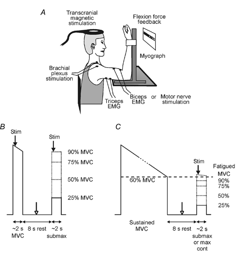
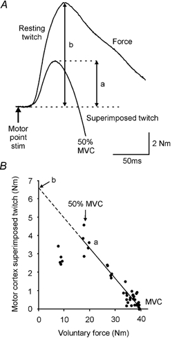
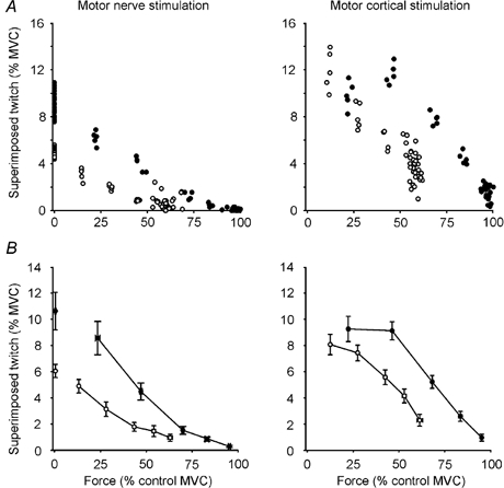
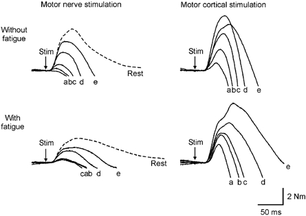
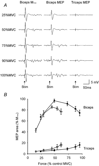
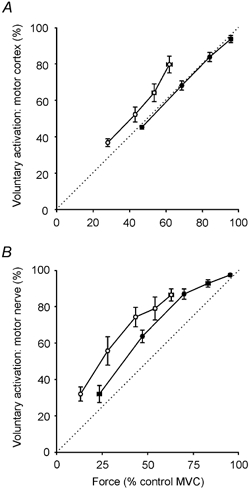
Recently, transcranial magnetic stimulation of the motor cortex (TMS) revealed impaired voluntary activation of muscles during maximal efforts. Hence, we evaluated its use as a measure of voluntary activation over a range of contraction strengths in both fresh and fatigued muscles, and compared it with standard twitch interpolation using nerve stimulation. Subjects contracted the elbow flexors isometrically while force and EMG from biceps and triceps were recorded. In one study, eight subjects made submaximal and maximal test contractions with rests to minimise fatigue. In the second study, eight subjects made sustained maximal contractions to reduce force to 60 % of the initial value, followed by brief test contractions. Force responses were recorded following TMS or electrical stimulation of the biceps motor nerve. In other contractions, EMG responses to TMS (motor evoked potentials, MEPs) or to stimulation at the brachial plexus (maximal M waves, Mmax) were recorded. During contractions of 50 % maximum, TMS elicited large MEPs in biceps (> 90 % Mmax) which decreased in size (to approximately 70 % Mmax) with maximal efforts. This suggests that faster firing rates made some motor units effectively refractory. With fatigue, MEPs were also smaller but remained > 70 % Mmax for contractions of 50-100 % maximum. For fresh and fatigued muscle, the superimposed twitch evoked by motor nerve and motor cortex stimulation decreased with increasing contraction strength. For nerve stimulation the relation was curvilinear, and for TMS it was linear for contractions of 50-100 % maximum (r2 = 1.00). Voluntary activation was derived using the expression: (1 - superimposed twitch/resting twitch) x 100. The resting twitch was measured directly for nerve stimulation and for TMS, it was estimated by extrapolation of the linear regression between the twitch and voluntary force. For cortical stimulation, this resulted in a highly linear relation between voluntary activation and force. Furthermore, the estimated activation corresponded well with contraction strength. Using TMS or nerve stimulation, voluntary activation was high during maximal efforts of fresh muscle. With fatigue, both measures revealed reduced voluntary activation (i.e. central fatigue) during maximal efforts. Measured with TMS, this central fatigue accounted for one-quarter of the fall in maximal voluntary force. We conclude that TMS can quantify voluntary activation for fresh or fatigued muscles at forces of 50-100 % maximum. Unlike standard twitch interpolation of the elbow flexors, voluntary activation measured with TMS varies in proportion to voluntary force, it reveals when extra output is available from the motor cortex to increase force, and it elicits force from all relevant synergist muscles.


Figures

Figure 1
Figure 1. Experimental apparatus and study protocols
A, experimental apparatus. B, study 1 protocol. Subjects performed pairs of contractions involving a brief maximal voluntary contraction (MVC) followed by a brief submaximal contraction without fatigue. Transcranial magnetic stimulation of the motor cortex (TMS), electrical stimulation of the brachial plexus, or electrical stimulation of the biceps brachii motor nerve was delivered during each contraction (filled arrows). In some contraction pairs, electrical stimulation of the biceps brachii motor nerve was also delivered at rest between contractions (open arrow). C, study 2 protocol. Subjects performed pairs of contractions of the fatigued elbow flexor muscles. Each contraction pair involved a sustained MVC (maintained until maximal force had decreased to 60 % of maximal force without fatigue) followed by a brief maximal or submaximal contraction. Arrows indicate the timing of the stimuli.
Figure 2
Figure 2. Methods used for calculation of voluntary activation with motor nerve or motor cortex stimulation
A, schematic representation of elbow flexion force following motor nerve stimulation. Arrow indicates the timing of the stimulus. Voluntary force trace with a resting muscle twitch (b) and superimposed twitch (a) evoked by motor nerve stimulation. Background forces have been offset to allow comparison of the twitches. B, single subject data displaying the linear correlation between the amplitude of the superimposed twitch (a) evoked by motor cortical stimulation between 50 and 100 % of maximal voluntary force (MVC). The linear regression was extrapolated to the y-axis and the y-intercept was taken as the estimated amplitude of the resting twitch (b) evoked by motor cortical stimulation.
Figure 4
Figure 4. Single subject and group data
Single subject (A) and group data (B; means ± s.e.m.) showing the amplitude of the superimposed twitch evoked by motor nerve stimulation or TMS (motor cortical stimulation) at varying contraction strengths with fatigue (○) and without fatigue (•). All forces are shown as percentages of maximal voluntary force (MVC) of the unfatigued muscle. With fatigue, maximal voluntary force was reduced to 60 % MVC and submaximal contractions were targeted to 90, 75, 50 and 25 % of the reduced maximal force.
Figure 3
Figure 3. Responses to motor nerve and motor cortex stimulation, with and without muscle fatigue
Average traces of elbow flexion force from one subject following a single motor nerve stimulus or transcranial magnetic stimulus to the motor cortex (TMS) at varying contraction strengths (a, 100 % MVC; b, 90 % MVC; c, 75 % MVC; d, 50 % MVC; e, 25 % MVC), with and without fatigue. Twitches to motor nerve stimulation were also recorded with the muscle at rest (dashed lines). Traces have been offset to allow comparison of the twitches. MVC, maximal voluntary contraction.
Figure 5
Figure 5. EMG responses to motor nerve and motor cortical stimulation
A, single-subject data showing averaged EMG responses following peripheral nerve stimulation (Mmax) and motor cortical stimulation (MEP) for the unfatigued biceps and following motor cortical stimulation (MEP) for the triceps. B, group data (means ± s.e.m.) showing the area of the motor evoked potential (MEP; expressed as a percentage of Mmax, see Methods) in the biceps (circles) and triceps (squares) muscles at varying contraction strengths with fatigue (open symbols) and without fatigue (filled symbols). MVC, maximal voluntary contraction.
Figure 6
Figure 6. Relationship between voluntary force and measures of voluntary activation
Voluntary muscle activation (%; see Methods), calculated with the use of motor cortical (A) and motor nerve stimulation (B) at varying contraction strengths with fatigue (○) and without fatigue (•). For motor cortical stimulation, data are plotted for contractions of 50, 75, 90 and 100 % maximal voluntary force (MVC). For motor nerve stimulation, contractions of 25, 50, 75, 90 and 100 % MVC are plotted. All forces are plotted as percentages of the MVC of the unfatigued muscle although with fatigue, contraction targets were set in relation to the fatigued maximal voluntary force. The dotted line is the line of identity.

Similar articles

Cited by

References

    1. Allen GE, McKenzie DK, Gandevia SC. Twitch interpolation of the elbow flexor muscles at high forces. Muscle Nerve. 1998;21:318–328. - PubMed
    1. Awiszus F, Wahl B, Meinecke I. Influence of stimulus cross talk on results of the twitch-interpolation technique at the biceps brachii muscle. Muscle Nerve. 1997;20:1187–1190. - PubMed
    1. Behm DG, Whittle J, Button D, Power K. Intermuscle differences in activation. Muscle Nerve. 2002;25:236–243. - PubMed
    1. Belanger AY, McComas AJ. Extent of motor unit activation during effort. J Appl Physiol. 1981;51:1131–1135. - PubMed
    1. Bigland-Ritchie B, Jones DA, Hosking GP, Edwards RHT. Central and peripheral fatigue in sustained maximum voluntary contractions of human quadriceps muscle. Clin Sci Mol Med. 1978;54:609–614. - PubMed

Publication types

LinkOut - more resources