. 2003 Sep 30;100 Suppl 1(Suppl 1):11889-95.
doi: 10.1073/pnas.1834204100. Epub 2003 Aug 8.

Activation of Notch signaling pathway precedes heart regeneration in zebrafish

Affiliations

Activation of Notch signaling pathway precedes heart regeneration in zebrafish

Angel Raya et al. Proc Natl Acad Sci U S A. .

Abstract

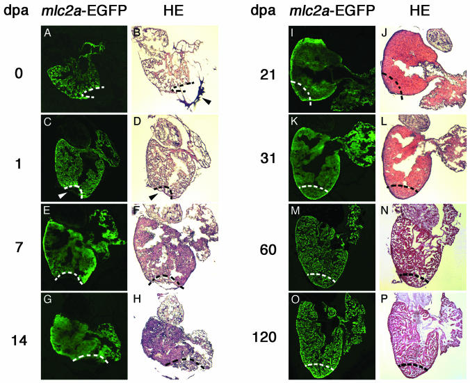
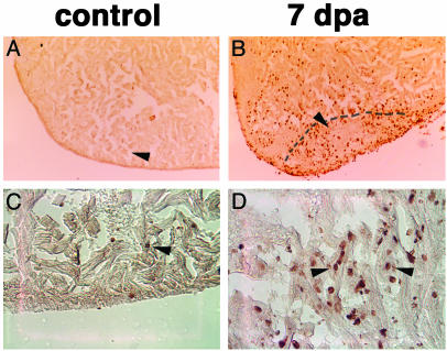
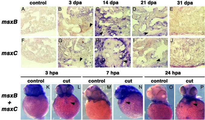
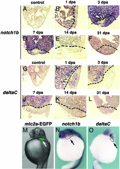
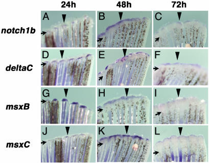
Several vertebrates display the ability to regenerate parts of their body after amputation. During this process, differentiated cells reenter the cell cycle and proliferate to generate a mass of undifferentiated cells. Repatterning mechanisms act on these cells to eventually shape a regenerated tissue or organ that replaces the amputated one. Experiments with regenerating limbs/fins in newts and zebrafish have shown that members of the Msx family of homeodomain-containing transcription factors play key roles during blastema formation and patterning. Here we show that adult zebrafish have a remarkable capacity to regenerate the heart in a process that involves up-regulation of msxB and msxC genes. We present evidence indicating that heart regeneration involves the execution of a specific genetic program, rather than redeployment of a cardiac development program. Preceding Msx activation, there is a marked increase in the expression of notch1b and deltaC, which we show are also up-regulated during fin regeneration. These data suggest a role for the Notch pathway in the activation of the regenerative response. Taken together, our results underscore the use of zebrafish as a model for investigating the process of regeneration in particular and the biology of stem cells in general. Advances in these fields will undoubtedly aid in the implementation of strategies for regenerative medicine.


Figures

Fig. 1.
Fig. 1.
Visualization of heart regeneration in mlc2a-EGFP transgenic zebrafish. Zebrafish heart regeneration was monitored by examining mlc2a-EGFP expression (A, C, E, G, I, K, M, and O) and histological characteristics of hematoxylin/eosin (HE)-stained sections (B, D, F, H, J, L, N, and P) for 0 (A and B),1(C and D),7(E and F), 14 (G and H), 21 (I and J), 31 (K and L), 60 (M and N), and 120 (O and P) dpa. Immediately after amputation, a blood clot forms near the open wound (arrowhead in B). By 1 dpa, this region has organized into a dense fibrin clot (arrowhead in D) devoid of EGFP expression (arrowhead in C). Progressive invasion of the fibrin clot by mlc2a-EGFP-positive cells occurs (E, G, I, and K), such that by 31 dpa, the amputated myocardium is completely replaced by regenerated tissue (K). Shown are sagittal sections through the midventricle. Dotted lines mark the amputation plane in A-H and the estimated amputation plane in I-P, for comparison.
Fig. 2.
Fig. 2.
Proliferation of cardiomyocytes is associated with zebrafish heart regeneration. Control (A and C) and amputated (B and D) zebrafish were labeled with BrdUrd for 7 d to detect cells undergoing mitosis during heart regeneration. Although an increase in BrdUrd incorporation occurs throughout the amputated hearts, a significant accumulation is observed near the amputation plane and in the tissue immediately beneath the regenerated epicardium (B). Arrowheads in A and B point to the area examined at higher magnification (×400) in C and D. We could identify BrdUrd-positive cells displaying morphological characteristics of cardiomyocytes (arrowhead in C; arrowheads and neighboring cells in D). Shown are sagittal sections through the midventricle stained for BrdUrd and counterstained with eosin. Dotted line marks the amputation plane.
Fig. 3.
Fig. 3.
Markers of early cardiac development are not up-regulated during zebrafish heart regeneration. Low levels of nkx2.5 expression are present throughout the myocardium of adult fish (A). However, no significant alterations in the expression of nkx2.5 are observed in regenerating hearts 7 (D), 14 (G), or 21 (J) dpa. Areas shown in D, G, and J illustrate the boundary between nonamputated myocardium and de novo formed (i.e., regenerated) tissue. Expression of the CARP-EGFP transgene, which is observed in the hearts of 24- (M), 36- (N), and 72- (O) hpf developing embryos but not adult control hearts (B) is also not induced during heart regeneration 7 (E), 14 (H), or 21 (K) dpa. Heart regeneration was confirmed in sections consecutive to those shown in B, E, H, and K by analyzing the expression of ventricular myosin heavy chain (vmhc) in control (C) and at 7 (F), 14 (I), or 21 (L) dpa. Shown are sagittal sections through the midventricle of adult hearts. Dotted lines mark the amputation plane. Sections in B, E, H, and K were overexposed (five times longer than required for the EGFP images in Fig. 1) so the background tissue autofluorescence could be seen.
Fig. 4.
Fig. 4.
msxC and msxB are expressed in the regenerating zebrafish heart. (A-J) The expression of msxB (A-E) and msxC (F-J) was analyzed in control adult hearts (A and F)and3(B and G), 14 (C and H), 21 (D and I), and 31 (E and J) dpa. At 3 dpa, both genes are up-regulated in myocardial tissue surrounding the lesion area (B and G) and reach a peak of expression by 14 dpa (C and H). Transcripts are no longer detected when heart regeneration is complete at 31 dpa (E and J). Shown are sagittal sections through the midventricle of adult hearts. Areas shown in B-E and G-J illustrate the boundary between nonamputated myocardium and de novo formed (i.e., regenerated) tissue. Arrowheads point to fibrin clot remnants in the lesion area. (K-P) Neither msxB nor msxC is expressed in the hearts of 24- to 48-hpf embryos (K, M, and O). However, both genes are dramatically up-regulated 3 (L), 7 (N), and 24 (P) h after removal of ≈50% of the developing atrium. Embryo views are frontal, anterior to the top. Arrowheads mark gene expression in the damaged embryonic hearts.
Fig. 5.
Fig. 5.
Up-regulation of notch1b and deltaC during zebrafish heart regeneration. (A-L) Expression of notch1b (A-F) and deltaC (G-L) was monitored in control adult hearts (A and G)and1(B and H),3(C and I),7(D and J), 14 (E and K) and 31 (F and L) dpa. Only weak expression of notch1b is detected in the ventricle of control adult hearts (A) but is dramatically up-regulated by 1 dpa (B) and persists until 3 dpa (C). By 14 dpa (C), expression of notch1b returns to control levels (E and F). Similarly, weak expression of deltaC is observed throughout the ventricle of control adult hearts (G), in a pattern similar to notch1b, but is dramatically up-regulated within 1 dpa (H) and returns to control levels after 7 dpa (J-L). Shown are sagittal sections through the midventricle. Dotted lines mark the amputation plane. (M-O) In contrast to regenerating adult hearts, neither notch1b (N) nor deltac (O) is expressed in the developing hearts of 24-hpf embryos. For comparison, M shows the location of the 24-hpf embryonic heart, as monitored by expression of an mlc2a-EGFP transgene. Arrows indicate the location of the embryonic heart (outlined in N and O). Embryo views are frontal, anterior to the top.
Fig. 6.
Fig. 6.
notch1b, deltaC, msxB, and msxC are expressed during caudal fin regeneration. Expression of notch1b (A-C), deltaC (D-F), msxB (G-I), and msxC (J-L) were analyzed after 24 (A, D, G, and J), 48 (B, E, H, and K), or 72 (C, F, I, and L) h postamputation of caudal fins. Both msxB and msxC have been reported to be expressed during fin regeneration (7) and are shown for comparison. notch1b is expressed in the blastema formation stage (24 h postamputation) (A), and the signal becomes prominent in the distal blastema 48 h postamputation (B) and is down-regulated at 72 h postamputation (C). deltaC is expressed in the blastema at 24 h postamputation (D) and is detected in the distal blastema at 48 (E) and 72 (F) h postamputation. Both msxB (G) and msxC (J) are expressed in the blastema at 24 h postamputation. The signals are detected in the distal blastema 48 h postamputation (H and K) and are down-regulated and more distally restricted at 72 h postamputation (I and L). Arrows indicate the level of amputation, and arrowheads point to representative gene expression.

Similar articles

Cited by

References

    1. Tsonis, P. A. (2000) Dev. Biol. 221, 273-284. - PubMed
    1. Brockes, J. P. & Kumar, A. (2002) Nat. Rev. Mol. Cell Biol. 3, 566-574. - PubMed
    1. Brockes, J. P. (1997) Science 276, 81-87. - PubMed
    1. Bryant, S. V., Endo, T. & Gardiner, D. M. (2002) Int. J. Dev. Biol. 46, 887-896. - PubMed
    1. Gardiner, D. M. & Bryant, S. V. (1996) Int. J. Dev. Biol. 40, 797-805. - PubMed

Publication types

LinkOut - more resources