. 2003 Sep 30;100 Suppl 1(Suppl 1):11896-903.
doi: 10.1073/pnas.1734139100. Epub 2003 Aug 8.

In vivo regeneration of murine prostate from dissociated cell populations of postnatal epithelia and urogenital sinus mesenchyme

Affiliations

In vivo regeneration of murine prostate from dissociated cell populations of postnatal epithelia and urogenital sinus mesenchyme

Li Xin et al. Proc Natl Acad Sci U S A. .

Abstract

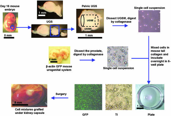
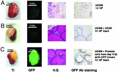
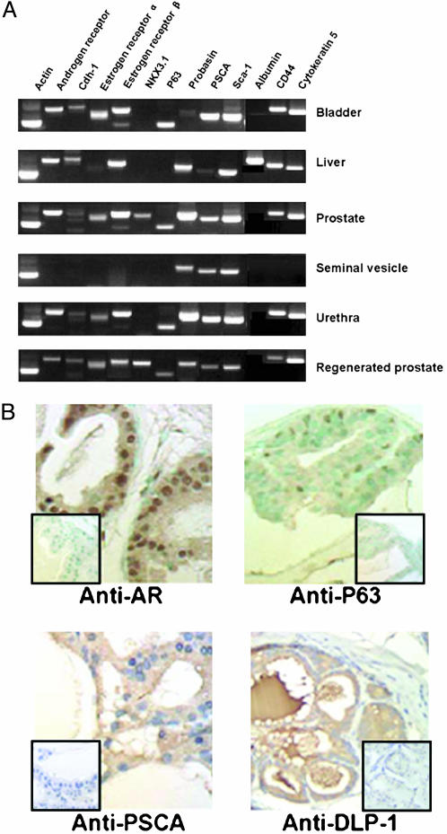
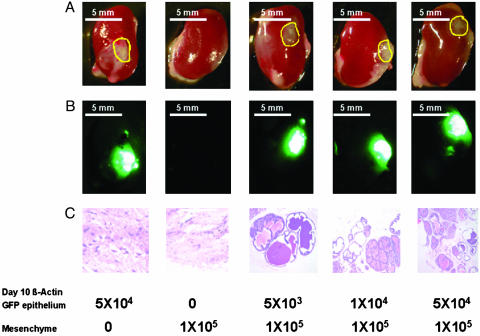
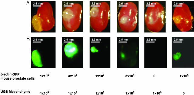
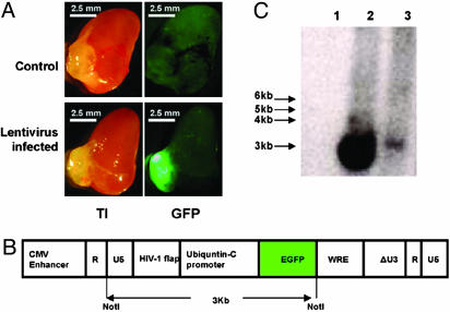
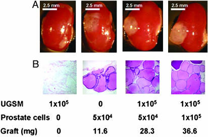
The existence of a postnatal prostate stem cell is supported by several types of evidence. Withdrawal of androgen leads to involution of the gland, but readdition can rapidly stimulate regeneration. Tissue fragments derived from mouse or rat prostatic epithelia from midgestation embryos or adult mice, when combined with tissue fragments from urogenital sinus mesenchyme and grafted under the kidney capsule, can regenerate prostatic structures. Indirect evidence supports that the stem cell population is contained within the basal layer. Purified prostatic stem cell preparations would be useful to define the physical and functional properties required for regeneration and to compare with cells that accumulate during abnormal growth states, like prostate cancer. We have developed a regeneration system using dissociated cell populations of postnatal prostate epithelia and embryonic urogenital sinus mesenchyme. Efficient in vivo regeneration of prostatic structures in the subcapsular space of the kidney was observed within 4-8 wk with as few as 103 epithelial cells from prostates derived from donors 10 d to 6 wk of age. The regenerated structures show a branching tubular epithelial morphology, with expression of a panel of markers consistent with prostate development. Donor epithelial populations can be readily infected with GFP expressing lentiviral vectors to provide integration markers and easy visualization. The cell preparations of urogenital sinus mesenchyme can be expanded in short-term in vitro culture while their inductive capabilities are retained. Further definition of the subpopulation of prostate epithelial cells containing the regeneration activity should be possible with such technologies.


Figures

Fig. 1.
Fig. 1.
Outline of the prostate regeneration system. Mid-UGS was dissected from day-16 C57BL/6 mouse embryos. Pelvic UGS was collected as described in Materials and Methods and trypsinized for 90 min in 4°C with 1% trypsin (Invitrogen), and UGSM was peeled off from the UGSE under a dissecting microscope. UGSM was digested with 0.8 mg/ml collagenase (GIBCO, 226 units/mg) in 10 ml of DMEM 10% FBS for 90 min at 37°C to produce single-cell populations. Prostate tissues were dissected from β-actin GFP mice, blade minced, and collagenase digested as for UGSM to produce single-cell suspensions. Varying numbers of UGSM cells and prostate cells were mixed in 40-50 μl of 1% rat-tail collagen (40) and incubated in a six-well plate in Bfs media (see Materials and Methods) at 37°C overnight (Plate). Under the transilluminating microscope (TI), the mixed cells were confirmed to be single-cell populations, whereas under the fluorescent microscope, a subportion was green. The next day, the collagen gel was implanted under the kidney capsule of an adult male CB.17SCID/SCID mouse. A testosterone pellet (12.5 mg per pellet, 90-d release, Innovative Research of America) was s.c. implanted at the same time. Refer to Materials and Methods for more details of reagents and procedures. Tissue photos were taken with a Leica MZFLIII dissecting microscope (×10). Bar sizes are indicated in each picture. Cell photos were taken with a Nikon Diaphot phase contrast microscope (×100).
Fig. 2.
Fig. 2.
Dissociated prostate epithelial cells combined with UGSM can regenerate the prostatic-like tissue. UGSE (1 × 10⁵) or 1 × 10⁵ 10-d-old β-actin GFP mouse prostate epithelial cells were mixed with 1 × 10⁵ UGSM, and the regeneration protocol was carried out as described in Materials and Methods and Fig. 1. Mice were killed 3 mo after the surgery, and regenerated tissues from the kidney capsules were collected. (A) UGSM (1 × 10⁵) alone was used as a negative control. (B) Graft initiated with 1 × 10⁵ UGSM and 1 × 10⁵ UGSE. (C) Graft composed of 1 × 10⁵ UGSM and 1 × 10⁵ dissociated 10-d-old β-actin GFP prostate epithelial cells. (A and B) From left to right: transilluminated image (TI) and green fluorescent image (GFP) of regenerated grafts; hematoxylin/eosin staining (H.E.), and the GFP antibody staining (GFP Ab staining) of regenerated tissue sections. GFP protein was detected by immunohistochemistry with rabbit polyclonal antibody against GFP (Abcam, 1:300 dilution) with the Envision+ system (DAKO). Photos were taken with a Leica MZFLIII dissecting microscope (×10). (Bars = 5 mm.)
Fig. 3.
Fig. 3.
Regenerated prostate has the same gene expression profile as normal prostate. (A) RT-PCR analysis of RNAs from regenerated prostate, 4-wk-old C57/BL6 mouse prostate, bladder, seminal vesicle, urethra, and liver. Gene expression analysis of different tissues for the indicated genes was performed by RT-PCR. RNA preparation, RT-PCR conditions, product length, and primer sequences are described in Materials and Methods. Actin was amplified to test the quality of the RNAs; albumin is a hepatocyte marker protein, and other proteins were previously reported to be expressed in prostate tissue (10, 42, 44). Note that NKX3.1 is detected only in the prostate as well as regenerated prostate. Sca-1, stem cell antigen-1. (B) Immunohistochemistry analysis of the expression of androgen receptor, P63, PSCA, and DLP-1 in regenerated prostate tissue. Tissue sections from regenerated prostate were stained with the polyclonal rabbit anti-androgen receptor (Santa Cruz Biotechnology, 1:200 dilution), polyclonal antiserum anti-secretions of mouse dorsolateral prostate (1:5,000 dilution) (43), or monoclonal hamster anti-PSCA, 2 μg/ml (44). Subsequently, sections were processed for immunohistochemistry by using the EnVision+ system (DAKO) or the ABC system (DAKO). P63 was detected with polyclonal mouse anti-mouse p63 (4A4, Santa Cruz Biotechnology, 1:200 dilution) and an ARK peroxidase kit (DAKO). (Inset) Lack of staining when using a corresponding control isotype IgG. (×400).
Fig. 4.
Fig. 4.
Number of postnatal day 10 epithelial cells required for effective regeneration. UGSM cells (1 × 10⁵) were mixed with 10-d-old β-actin GFP mouse prostate epithelial cells ranging from 5 × 10³ to 5 × 10⁴, and the regeneration protocol was carried out as described in Materials and Methods and Fig. 1. Mice were killed 1 mo after surgery, and regenerated tissues from the kidney capsules were collected. UGSM alone and β-actin GFP mouse prostate epithelial cells alone were used as controls. (A) Transillumination. (B) Green fluorescence image of the regenerated tissues under kidney capsules. (C) Hematoxylin/eosin staining of the regenerated tissue sections. Numbers below the panels indicate the cell number of the UGSM and the prostate epithelial cells. Yellow circles in A show the positions of the regenerated tissues. A and B photos were taken with a Leica MZFLIII dissecting microscope (×10). (Bars = 5 mm.) (C) Photo was taken with a Nikon Diaphot phase contrast microscope (×100).
Fig. 5.
Fig. 5.
Regeneration by using adult mice prostate epithelial cells. UGSM cells (1 × 10⁵) were mixed with 6-wk-old β-actin GFP mouse prostate epithelial cells ranging from 3 × 10³ to 1 × 10⁵, and the regeneration experiment was done as described in Materials and Methods. Grafts composed solely of UGSM or UGSE cells were used as controls. Mice were killed 2 mo after surgery, and regenerated tissues under the kidney capsules were collected. Transillumination (A) and green fluorescence (B) images of the regenerated tissues under the kidney capsule. Numbers below the panels indicate the cell number of UGSM and prostate epithelial cells. Photos were taken with a Leica MZFLIII dissecting microscope (×10). (Bars = 2.5 mm.)
Fig. 6.
Fig. 6.
Prostate regeneration with GFP-lentivirus-infected prostate epithelial cells. Six-week-old C57/BL6 mouse prostate epithelial cells were infected by GFP-expressing lentivirus stock as described in Materials and Methods. Six-week-old prostate epithelial cells (1 × 10⁵) infected by GFP-lentivirus or control cells were mixed with 1 × 10⁵ UGSM cells and processed for regeneration as described in Materials and Methods and Fig. 1. Mice were killed 2 mo after surgery, and the regenerated tissues from the kidney capsules were collected. (A) Transillumination (TI, Left) and green fluorescence (GFP, Right) images of regenerated prostate using uninfected prostate epithelial cells (Control, Upper) and regenerated prostate using GFP-lentivirus infected prostate epithelial cells (Lentivirus infected, Lower). Photos were taken with a Leica MZFLIII dissecting microscope (×10). (Bars = 2.5 mm.) (B) Schematic diagram of GFP-lentivirus genomic backbone. The expression of GFP is driven by the ubiquitin-C promoter (36, 37). On NotI digestion, a 3-kb band is released from the lentivirus backbone (diagram adapted from ref. 36). (C) Southern blot analysis of the integration of lentivirus in the regenerated prostate. A 3-kb band can be released on NotI digestion of the genomic DNA. Southern blot was performed as described in Materials and Methods. NIH/3T3 cells infected with GFP-lentivirus were used as positive control. A band of ≈3 kb was detected in the positive control (lane 2, 15 μg of genomic DNA) and the regenerated prostate by using GFP-lentivirus infected prostate epithelial cells (lane 3, 10 μg of genomic DNA) but not in the regenerated prostate by using uninfected epithelial cells (lane 1, 10 μg of genomic DNA).
Fig. 7.
Fig. 7.
In vitro cultured UGSM cells are able to regenerate prostate. UGSM cells were cultured in Bfs media as described in Materials and Methods for 1 wk. Four-week-old prostate epithelial cells (1 × 10⁵ or 5 × 10⁴) were mixed with 1 × 10⁵ UGSM, and the regeneration protocol was done as described in Materials and Methods. Grafts composed of UGSM only or epithelial cells were used as controls. Mice were killed 2 mo after surgery, and regenerated tissues under the kidney capsules were collected. (A) Transillumination image of regenerated prostate under kidney capsule. (B) Hematoxylin/eosin staining of regenerated tissue sections. Numbers below the panels indicate the number of the UGSM cells and the prostate epithelial cells used and the weight of the grafts. Note that the graft composed of only epithelial cells grew to a small extent compared with the prostate regenerated from UGS and epithelial cells; its weight and size are much smaller, and the number of branching tubular epithelial structures is lower. (A) Photos were taken with a Leica MZFLIII dissecting microscope (×10). (Bars = 2.5 mm.) (B) Photos were taken with a Nikon Diaphot phase contrast microscope (×100).

References

    1. Weissman, I. L., Anderson, D. J. & Gage, F. (2001) Annu. Rev. Cell Dev. Biol. 17, 387-403. - PubMed
    1. Morrison, S. J., Hemmati, H. D., Wandycz, A. M. & Weissman, I. L. (1995) Proc. Natl. Acad. Sci. USA 92, 10302-10306. - PMC - PubMed
    1. Jackson, K. A., Majka, S. M., Wulf, G. G. & Goodell, M. A. (2002) J. Cell Biochem. 38, Suppl., 1-6. - PubMed
    1. Reya, T., Morrison, S. J., Clarke, M. F. & Weissman, I. L. (2001) Nature 414, 105-111. - PubMed
    1. Ramalho-Santos, M., Yoon, S., Matsuzaki, Y., Mulligan, R. C. & Melton, D. A. (2002) Science 298, 597-600. - PubMed

Publication types

MeSH terms