. 2003 Aug 19;100(17):9974-9.
doi: 10.1073/pnas.1732638100. Epub 2003 Aug 8.

Defective double-strand DNA break repair and chromosomal translocations by MYC overexpression

Affiliations

Defective double-strand DNA break repair and chromosomal translocations by MYC overexpression

Asa Karlsson et al. Proc Natl Acad Sci U S A. .

Abstract

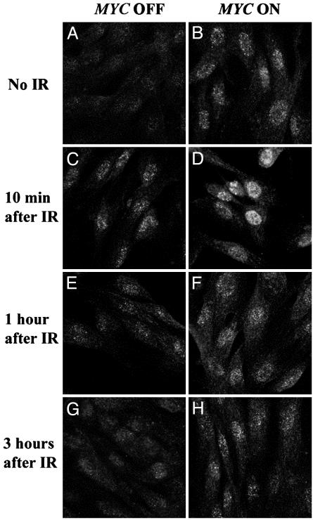
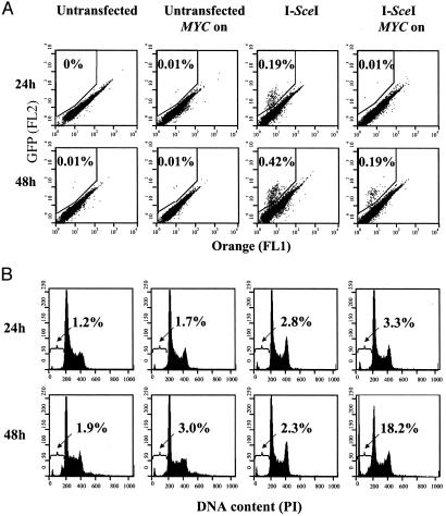
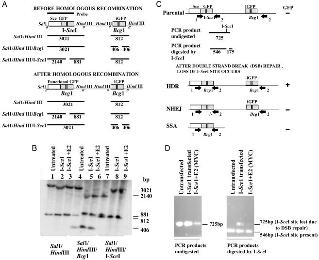
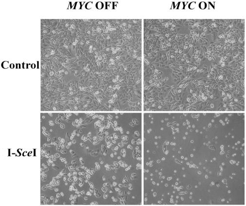
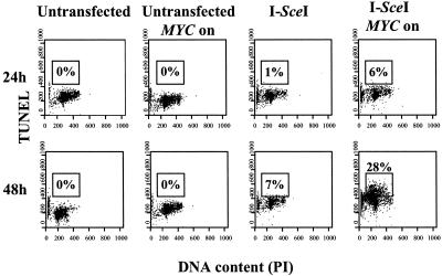
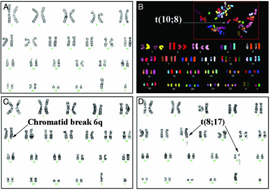
DNA repair mechanisms are essential for the maintenance of genomic integrity. Disruption of gene products responsible for DNA repair can result in chromosomal damage. Improperly repaired chromosomal damage can result in the loss of chromosomes or the generation of chromosomal deletions or translocations, which can lead to tumorigenesis. The MYC protooncogene is a transcription factor whose overexpression is frequently associated with human neoplasia. MYC has not been previously implicated in a role in DNA repair. Here we report that the overexpression of MYC disrupts the repair of double-strand DNA breaks, resulting in a several-magnitude increase in chromosomal breaks and translocations. We found that MYC inhibited the repair of gamma irradiation DNA breaks in normal human cells and blocked the repair of a single double-strand break engineered to occur in an immortal cell line. By spectral karyotypic analysis, we found that MYC even within one cell division cycle resulted in a several-magnitude increase in the frequency of chromosomal breaks and translocations in normal human cells. Hence, MYC overexpression may be a previously undescribed example of a dominant mutator that may fuel tumorigenesis by inducing chromosomal damage.


Figures

Fig. 1.
Fig. 1.
Overexpression of MYC induces formation of γ-H2AX foci in normal human fibroblasts. NHF-MYCER cells were stained for γ-H2AX (green) after 2 h without (A) and with (B) MYC activation and analyzed by confocal microscopy. NHF-MYCER cells without (C, E, and G) and with (D, F, and H) MYC activation were γ-irradiated and stained for formation of γ-H2AX-foci 10 min (C and D), 1h(E and F),and3h(G and H) after irradiation (IR). Representative data from one of three experiments are shown.
Fig. 2.
Fig. 2.
MYC overexpression prevents the repair of a DSB through homologous recombination and induces apotosis. DRAA8-MYCER cells were untreated or electroporated with I-SceI in the presence or absence of E2 and analyzed 24 or 48 h later. (A) GFP expression was measured by flow cytometry to determine the frequency of cells that successfully underwent homologous recombination. (B) Propodium iodide staining of cells was analyzed by flow cytometry to measure apoptosis. The percentage of cells that had undergone homologous recombination or were undergoing apoptosis is shown. FL1, green fluorescence; FL2, orange fluorescence. Representative data from one of three experiments are shown.
Fig. 3.
Fig. 3.
MYC overexpression globally suppresses DNA repair of DSBs. (A) Map of the DR-GFP substrate before and after homologous recombination. Sizes of expected fragments are indicated in base pairs. (B) Southern analysis confirmed that the GFP-positive cells have undergone repair of their DSBs through short-track gene conversion or homologous recombination. Genomic DNA from DRAA8-MYCER cells that were untransfected and GFP-positive cells that were I-SceI-transfected in the presence or absence of MYC activation with E2 was digested with the indicated restriction enzymes and analyzed by Southern analysis. The 812-bp band in lane 4 is due to incomplete digestion by Bcg1. (C) DSB repair mediated through HDR, NHEJ, and SSA results in the loss of the I-SceI site. The PCR-based assay using genomic DNA and primers 1 and 2 (arrow) results in a 725-bp fragment, which can then be cleaved by I-SceI to yield 546- and 175-bp fragments. After I-SceI transfection and DSB repair, the PCR product can no longer be cleaved by I-SceI due to loss of the I-SceI site. (D) Agarose gel analysis of the PCR products from the untransfected, I-SceI-transfected, and I-SceI-transfected and MYC activated cells were either digested by I-SceI (Right) or not (Left). In all three cell lines, the PCR product was a 725-bp fragment (Left). When the PCR products were digested by I-SceI (Right), the untransfected cell line yielded a 546-bp fragment and a 175-bp fragment that is not shown; I-SceI-transfected cells yielded 546- and 725-bp fragments caused by DSB repair, whereas I-SceI-transfected and MYC activated cells were found to yield only a 546-bp fragment due to impairment of DSB repair.
Fig. 4.
Fig. 4.
Cell death caused by MYC overexpression in the presence of a DSB. DRAA8-MYCER cells with or without MYC activation were electroporated with I-SceI, cultured for 48 h, then examined by phase microscopy. Over 90% of the I-SceI-transfected cells overexpressing MYC have died, as shown by the low cell density and large number of floating cells. I-SceI-transfected cells not overexpressing MYC grow at a higher cell density and do not show a large number of floating cells. Untransfected cells were included as a control. Representative data from one of three experiments are shown.
Fig. 5.
Fig. 5.
MYC overexpression impairs DSB repair and induces apoptosis. DRAA8-MYCER cells with or without MYC activation were electroporated with I-SceI, cultured for the indicated times, then analyzed by flow cytometry for DNA content by propidium iodide staining and DSB by TUNEL. The percentage of cells that are TUNEL-positive is indicated. Representative data from one of at least three experiments are shown.
Fig. 6.
Fig. 6.
Overexpression of MYC induces chromosomal breaks and translocations. (A) Normal karyotype of NHF-MYCER in the absence of E2. Shown is nonreciprocal translocation (B), t(10, 8) by SKY and chromatid break (C), and reciprocal chromosomal translocation, t(8, 17)(q24.3;q21) (D) by routine karyotypic analysis in NHF-MYCER after 48 h of MYC activation. Representative metaphases were chosen.

References

    1. Zhou, B. B. & Elledge, S. J. (2000) Nature 408, 433–439. - PubMed
    1. Khanna, K. K. & Jackson, S. P. (2001) Nat. Genet. 27, 247–254. - PubMed
    1. Pierce, A. J., Stark, J. M., Araujo, F. D., Moynahan, M. E., Berwick, M. & Jasin, M. (2001) Trends Cell. Biol. 11, S52–S59. - PubMed
    1. Myung, K., Chen, C. & Kolodner, R. D. (2001) Nature 411, 1073–1076. - PubMed
    1. Weiss, R. S., Matsuoka, S., Elledge, S. J. & Leder, P. (2002) Curr. Biol. 12, 73–77. - PubMed

Publication types