. 2003 Aug 19;100(17):9968-73.
doi: 10.1073/pnas.1631086100. Epub 2003 Aug 8.

The 15-lipoxygenase-1 product 13-S-hydroxyoctadecadienoic acid down-regulates PPAR-delta to induce apoptosis in colorectal cancer cells

Affiliations

The 15-lipoxygenase-1 product 13-S-hydroxyoctadecadienoic acid down-regulates PPAR-delta to induce apoptosis in colorectal cancer cells

Imad Shureiqi et al. Proc Natl Acad Sci U S A. .

Abstract

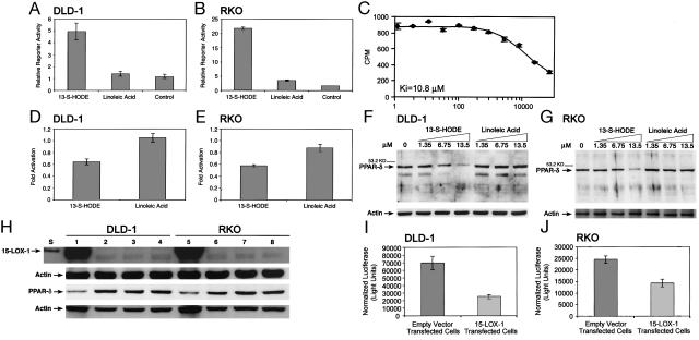
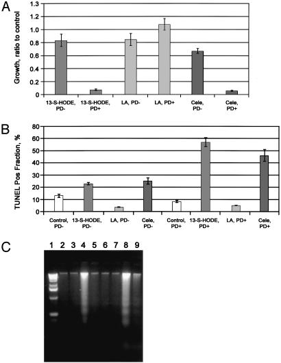
Diminished apoptosis, a critical event in tumorigenesis, is linked to down-regulated 15-lipoxygenase-1 (15-LOX-1) expression in colorectal cancer cells. 13-S-hydroxyoctadecadienoic acid (13-S-HODE), which is the primary product of 15-LOX-1 metabolism of linoleic acid, restores apoptosis. Nonsteroidal antiinflammatory drugs (NSAIDs) transcriptionally up-regulate 15-LOX-1 expression to induce apoptosis. Peroxisome proliferator-activated receptors (PPARs) are nuclear receptors for linoleic and arachidonic acid metabolites. PPAR-delta promotes colonic tumorigenesis. NSAIDs suppress PPAR-delta activity in colon cancer cells. The mechanistic relationship between 15-LOX-1 and PPAR-delta was previously unknown. Our current study shows that (i) 13-S-HODE binds to PPAR-delta, decreases PPAR-delta activation, and down-regulates PPAR-delta expression in colorectal cancer cells; (ii) the induction of 15-LOX-1 expression is a critical step in NSAID down-regulation of PPAR-delta and the resultant induction of apoptosis; and (iii) PPAR-delta is an important signaling receptor for 13-S-HODE-induced apoptosis. The in vivo relevance of these mechanistic findings was demonstrated in our tumorigenesis studies in nude mouse xenograft models. Our findings indicate that the down-regulation of PPAR-delta by 15-LOX-1 through 13-S-HODE is an apoptotic signaling pathway that is activated by NSAIDs.


Figures

Fig. 1.
Fig. 1.
Effects of 15-LOX-1 and 13-S-HODE on PPAR-δ expression and activation in colorectal cancer cells. (A and B) Effects of 13-S-HODE on the PPAR-δ LBD in DLD-1 (A) and RKO (B) cells. DLD-1 and RKO cells were treated with 13.5 μM 13-S-HODE, linoleic acid, or an equivalent volume of vehicle solvent (DMSO, control). Luciferase activity for the Gal4-PPAR-δ LBD and pMH-100-TK-luc reporter plasmid system was measured and normalized to β-gal activity (relative to 100,000 β-gal units). Values shown are the means and SEMs of triplicate experiments. (C) Scintillation proximity assay of 13-S-HODE binding to PPAR-δ. Values on x axis are nM 13-S-HODE. (D and E) Effects of 13-S-HODE (13.5μM) on PPAR-δ activation. Luciferase activity for the DRE-luciferase reporter vector was measured and normalized toβ-gal activity (relative to 100,000 β-gal units). Values shown are the fold activation relative to control (DMSO-treated cells) and represent the means of triplicate experiments. Error bars represent SEM. (F and G) Effects of 13-S-HODE on PPAR-δ expression. Western blot analyses for PPAR-δ expression in cells treated with 13-S-HODE or linoleic acid, cultured for 24 h, and then harvested; repeated experiments showed similar results. Control cells (0) were treated with DMSO only. Equal loading was assessed by probing for actin. (H) Effects of 15-LOX-1 expression on PPAR-δ expression. RKO and DLD-1 cells were transfected with 15-LOX-1 expression vector, empty vector, or transfection media alone (mock transfection). Western blots show the expression of 15-LOX-1 and PPAR-δ in cells harvested at 24 h. Similar results were observed at 48 h and with repeated experiments. Lanes: S, standard 15-LOX-1 recombinant protein; 1 and 5, 15-LOX-1 expression vector transfected cells; 2 and 6, empty vector-transfected cells; 3 and 7, mock transfections; 4 and 8, cells without transfections. (I and J) Effects of 15-LOX-1 expression on PPAR-δ activity. DLD-1 (I) and RKO (J) cells were transfected with 15-LOX-1 expression vector or the empty vector and with DRE-luciferase reporter and pSV-βgal vectors. Luciferase activity values were normalized toβ-gal activity. Shown values are means and SEMs from 48-h experiments. Repeated experiments at 24 and 48 h showed similar results. 15-LOX-1 expression reduced PPAR-δ activation significantly compared (t test) with empty-vector-transfected RKO (E-RKO) (P = 0.0125) and empty-vector-transfected DLD-1 (P = 0.0052) cells.
Fig. 2.
Fig. 2.
NSAID effects on PPAR-δ expression in colorectal cancer cells. (Top) Effects of NSAIDs on PPAR-δ RNA expression in colorectal cancer cells. Equal RNA loading was assessed by probing for GAPDH. (Middle) Time course for the effects of celecoxib on PPAR-δ protein expression in DLD-1 cells. Similar results were observed with RKO cells (data not shown). (Bottom) Effects of NSAIDs on PPAR-δ protein expression in RKO cells. Cells were treated with NSAIDs, harvested 72 h later, processed for Western blotting, and probed with PPAR-δ antibody. Lanes: 1, control; 2, NS-398; 3, indomethacin; 4, sulindac; 5, sulindac sulfone. Similar results were observed with DLD-1 cells (data not shown) and with repeated experiments.
Fig. 3.
Fig. 3.
Effects of celecoxib-induced 15-LOX-1 expression on PPAR-δ expression and apoptosis. (A) Effects of stable transfection of 15-LOX-1-AS on celecoxibinduced 15-LOX-1 in RKO cells. S, standard positive control of human 15-LOX-1 recombinant protein; –, control cells that were treated with DMSO (celecoxib solvent); +, celecoxib-treated cells; Empty vector, empty-vector-transfected cells; 1–4, AS-transfected clones. (B) Effects of celecoxib on 15-LOX-1 and PPAR-δ in stably transfected RKO cells with 15-LOX-1-AS clone 4 in protracted cell-culture exposure (240 h after celecoxib treatment). Similar results were observed with AS clone 3 (data not shown). (C) Effects of blocking 15-LOX-1-induced expression by celecoxib on growth inhibition by celecoxib. Growth ratios of celecoxib (cele)treated WT RKO (W-RKO), E-RKO, and 15-LOX-1-AS clone 4 stably transfected cells (15-LOX-1-AS) to control cells (DMSO-treated only) at 72 h. Mean ± SEM of triplicate experiments. (cele, 15-LOX-1-AS vs. cele, W-RKO, P = 0.0008; cele, 15-LOX-1-AS vs. cele, E-RKO, P = 0.0007; cele, W-RKO vs. cele, E-RKO, P = 0.88). (D) Effects of blocking 15-LOX-1 expression on celecoxib-induced apoptosis measured by terminal deoxynucleotidyltransferase-mediated dUTP nick end labeling (TUNEL) assay in RKO cells. Percentages of TUNEL-positive cells in W-RKO, E-RKO, and 15-LOX-1-AS cells are means ± SEM of triplicate experiments at 72 h after celecoxib or DMSO (control) treatment. (cele, 15-LOX-1-AS vs. cele, W-RKO, P < 0.0001; cele, 15-LOX-1-AS vs. cele, E-RKO, P < 0.0001). (E) Effects of blocking 15-LOX-1 expression on celecoxib-induced apoptosis as measured by sub-G1 fractions in RKO cells. Sub-G1 fractions in W-RKO, E-RKO, and 15-LOX-1-AS treated with either celecoxib or DMSO (control) and harvested 72 h later are means ± SEM of triplicate experiments. (cele, 15-LOX-1-AS vs. cele, W-RKO, P = 0.0015; cele, 15-LOX-1-AS vs. cele, E-RKO, P < 0.0001). (F) Effects of blocking 15-LOX-1 expression on celecoxib-induced apoptosis as measured by DNA laddering assay. Lanes: 1, standard DNA ladder; 2, W-RKO cells; 3, W-RKO cells treated with celecoxib; 4, E-RKO cells; 5, E-RKO cells treated with celecoxib; 6, 15-LOX-1-AS (control); 7, 15-LOX-1-AS cells treated with celecoxib.
Fig. 4.
Fig. 4.
PPAR-δ effects on celecoxib and 13-S-HODE-induced apoptosis in colon cancer cells. (A) Growth ratios of treated cells represent the number of viable cells divided by the number of viable cells in the control experiment (DMSO-treated only) at 72 h. PD+, WT HCT-116 cells that express PPAR-δ;PD–, HCT-116 cells that are PPAR-δ null. 13-S-HODE and linoleic acid (LA) concentrations were 13.5 μM. Means ± SEMs of triplicate experiments are shown. (13-S-HODE, PD–, vs. 13-S-HODE, PD+, P < 0.0001; celecoxib, PD–, vs. celecoxib, PD+, P < 0.003.) Repeated experiments showed similar results. (B) Effects of silencing PPAR-δ expression on apoptosis induction by 13-S-HODE and celecoxib, measured by TUNEL assay. Means ± SEMs of triplicate experiments are shown. (13-S-HODE, PD– vs. 13-S-HODE, PD+, P < 0.0001; celecoxib, PD– vs. celecoxib, PD+, P < 0.001; linoleic acid, PD– vs. linoleic acid, PD+, P = 0.672.) Similar results were found with sub-G1 fraction assays and in triplicate experiments (data not shown). (C) Effects (measured by DNA laddering) of silencing PPAR-δ expression on apoptosis induction by celecoxib and 13-S-HODE. Lanes: 1, standard DNA ladder; 2, PD+ cells treated with DMSO only (control); 3, PD– cells treated with DMSO only (control); 4, PD+ cells treated with 13-S-HODE; 5, PD– cells treated with 13-S-HODE; 6, PD+ cells treated with linoleic acid; 7, PD– cells treated with linoleic acid; 8, PD+ cells treated with celecoxib; 9, PD– cells treated with celecoxib.
Fig. 5.
Fig. 5.
Effects of celecoxib-induced 15-LOX-1 expression on PPAR-δ expression and tumorigenesis in vivo. Wild type RKO (W RKO) and RKO cells transfected with 15-LOX-1-AS clone 3 (15-LOX AS-3), 15-LOX-1-AS clone 4 (15-LOX AS-4), and E-RKO were grown as xenografts in nude mice. Animals were randomized to celecoxib treatment or a control diet. Celecoxib inhibited the growth of E-RKO (A) but not 15-LOX AS-3 (B) or 15-LOX AS-4 (C) cells. (D) 15-LOX-1 and PPAR-δ Western blot analyses of W-RKO, 15-LOX AS-3, and 15-LOX AS-4 tissue xenografts. Lanes: S, standard 15-LOX-1 recombinant protein; 1, W-RKO (WT); 2, W-RKO + celecoxib; 3, 15-LOX AS-3 (AS-3); 4, 15-LOX AS-3 + celecoxib; 5, 15-LOX AS-4 (AS-4); 6, 15-LOX AS-4 + celecoxib. Actin expression was used to assess equal loading. (E) Proposed model of 15-LOX-1, 13-S-HODE, and PPAR-δ as a signaling pathway that can be modulated by NSAIDs to induce apoptosis and inhibit colorectal tumorigenesis.

Similar articles

Cited by

References

    1. Hall, P. A., Coates, P. J., Ansari, B. & Hopwood, D. (1994) J. Cell Sci. 107, 3569–3577. - PubMed
    1. Evan, G. I. & Vousden, K. H. (2001) Nature 411, 342–348. - PubMed
    1. Tomlinson, I. P. & Bodmer, W. F. (1995) Proc. Natl. Acad. Sci. USA 92, 11130–11134. - PMC - PubMed
    1. Johnstone, R. W., Ruefli, A. A. & Lowe, S. W. (2002) Cell 108, 153–164. - PubMed
    1. Nicholson, D. W. (2000) Nature 407, 810–816. - PubMed

Publication types

MeSH terms

LinkOut - more resources