. 2003 Sep;52(9):1327-32.
doi: 10.1136/gut.52.9.1327.

Upregulation of lymphotoxin beta expression in liver progenitor (oval) cells in chronic hepatitis C

Affiliations

Upregulation of lymphotoxin beta expression in liver progenitor (oval) cells in chronic hepatitis C

K N Lowes et al. Gut. 2003 Sep.

Abstract

Background: Bipotent liver progenitor (oval) cells with the ability to differentiate into hepatocytes and biliary epithelium have recently been identified in human subjects with hepatitis C. Animal studies suggest that members of the tumour necrosis factor family, including lymphotoxin beta (LT-beta), regulate oval cell proliferation in liver disease, but its role in human liver disease is unclear.

Aims: This study seeks to establish a role for LT-beta in hepatitis C related liver injury and to provide evidence that its increased expression is related to the presence of oval cells.

Methods: Liver biopsy specimens were obtained from patients with chronic hepatitis C virus (HCV) infection (n=20). Control liver samples (n=5) were obtained from liver resection or transplant surgery. LT-beta expression in liver biopsy specimens was studied using quantitative real time polymerase chain reaction and immunohistochemistry.

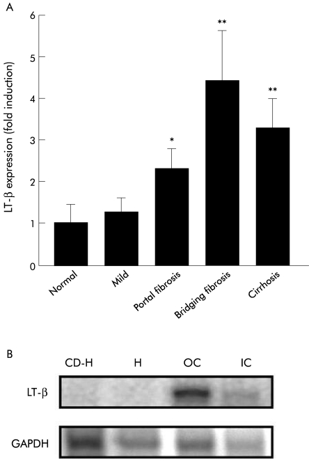
Results: LT-beta mRNA levels were similar in control and HCV liver in the absence of fibrosis. In subjects with portal fibrosis, LT-beta mRNA levels were elevated 2.2-fold over control liver levels (p=0.04). In subjects with bridging fibrosis, LT-beta mRNA levels increased 4.4-fold over control liver levels (p=0.02). LT-beta mRNA levels in subjects with established cirrhosis were increased 3.3-fold compared with controls and 2.6-fold compared with mild liver damage (p=0.02). Immunohistochemical analysis established that LT-beta was expressed by oval cells, inflammatory cells, and small portal hepatocytes.

Conclusions: In chronic HCV infection, LT-beta expression is observed in multiple hepatic cell types, including oval cells. LT-beta expression is significantly increased when fibrosis or cirrhosis is present, suggesting a role for LT-beta in the pathogenesis of chronic hepatitis C and a possible role in oval cell mediated liver regeneration.


Figures

Figure 1
Figure 1
(A) Expression of lymphotoxin β (LT-β) mRNA in liver in chronic hepatitis C. LT-β mRNA levels detected in liver biopsies from chronic hepatitis C patients increased significantly with fibrosis and cirrhosis. No significant difference was observed between control liver (n=5) and subjects with non-fibrotic liver damage (n=5). LT-β mRNA levels were 2.3-fold and 4.4-fold higher than control liver in subjects with portal fibrosis (n=5) and bridging fibrosis (n=5), respectively. LT-β mRNA levels were increased 3.3-fold in cirrhotic liver (n=5) compared with control liver. Results are expressed as mean (SEM) (*p=0.04, **p=0.02). (B) Ribonuclease protection analysis performed on RNA extracted from hepatocytes, oval cells, and inflammatory cells isolated from mice that received a choline deficient ethionine supplemented (CDE) diet for four weeks. LT-β mRNA is expressed by oval cells (O) and inflammatory cells (IC). LT-β mRNA was undetectable in normal hepatocytes (H) and CDE-treated hepatocytes (CD-H). GAPDH, glyceraldehyde-3-phosphate dehydrogenase.
Figure 2
Figure 2
Immunohistochemical staining illustrating cellular localisation of lymphotoxin β (LT-β) in human hepatitis C virus (HCV) infected liver with bridging fibrosis. (A–F) Serial staining for haematoxylin and eosin (H&E), leucocyte common antigen (LCA) (CD45), π-glutathione S-transferase (GST), M2-pyruvate kinase (M2-PK), proliferating cell nuclear antigen (PCNA), and LT-β suggests oval cells in HCV infected liver express LT-β. (A) H&E staining illustrating periportal cuffing by lymphocytes in chronic hepatitis C. The portal vein is labelled (pv). (B) LCA (CD45) expression illustrates that the mass of small cells clustered around the portal vein are CD45 positive. Oval cells (small arrows) were identified towards the periphery of this region by expression of π-GST (C) and M2-PK (D). (E) The small cells clustered directly around the portal vein are PCNA negative while oval-like cells (small arrows) are weakly PCNA positive and large centrally located hepatocytes (large arrow) are strongly PCNA positive. (F) LT-β expression was restricted to the periphery of the inflammatory cell mass, suggesting that the majority of LCA positive cells are not expressing LT-β. Cell types expressing LT-β include small portal hepatocytes (large arrow), oval cells (small arrows), and inflammatory cells (arrowhead). Bile ducts and large hepatocytes are negative. (G) Periportal cuffing by lymphocytes in chronic hepatitis C, portal fibrosis. This region contains LT-β positive oval cells (small arrows) and small hepatocytes adjacent to the portal vein with membranous and cytoplasmic LT-β staining (large arrow). LT-β expression is observed in some inflammatory cells (arrowhead). (H) Chronic hepatitis C with cirrhosis shows similar LT-β staining patterns, with oval cells (small arrows) and some inflammatory cells (arrowhead) staining positive for LT-β. Hepatocytes adjacent to fibrous septae also express LT-β (large arrow). Magnification bars represent 100 μm.

Similar articles

Cited by

References

    1. Lowes KN, Brennan BA, Yeoh GC, et al. Oval cell numbers in human chronic liver diseases are directly related to disease severity. Am J Pathol 1999;154:537–41. - PMC - PubMed
    1. Vessey CJ, Hall PM. Hepatic stem cells: a review. Pathology 2001;33:130–41. - PubMed
    1. Sell S. Heterogeneity and plasticity of hepatocyte lineage cells. Hepatology 2001;33:738–50. - PubMed
    1. Knight B, Yeoh GCT, Husk KL, et al. Impaired preneoplatic changes and liver tumour formation in tumour necrosis factor receptor type 1 knockout mice. J Exp Med 2000;192:1809–18. - PMC - PubMed
    1. Evarts RP, Hu Z, Fujio K, et al. Activation of the hepatic stem cell compartment in the rat: role of transforming growth factor, hepatocyte growth factor, and acidic fibroblast growth factor in early proliferation. Cell Growth Diff 1993;4:555–61. - PubMed

Publication types