. 2003 Sep;52(9):1347-54.
doi: 10.1136/gut.52.9.1347.

Vascular endothelial growth factor and receptor interaction is a prerequisite for murine hepatic fibrogenesis

Affiliations

Vascular endothelial growth factor and receptor interaction is a prerequisite for murine hepatic fibrogenesis

H Yoshiji et al. Gut. 2003 Sep.

Abstract

Background: It has been shown that expression of the potent angiogenic factor, vascular endothelial growth factor (VEGF), and its receptors, flt-1 (VEGFR-1) and KDR/Flk-1 (VEGFR-2), increased during the development of liver fibrosis.

Aims: To elucidate the in vivo role of interaction between VEGF and its receptors in liver fibrogenesis.

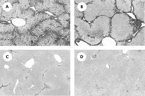
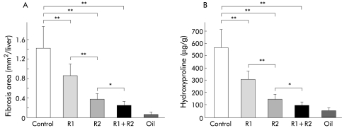
Methods: A model of CCl(4) induced hepatic fibrosis was used to assess the role of VEGFR-1 and VEGFR-2 by means of specific neutralising monoclonal antibodies (R-1mAb and R-2mAb, respectively). R-1mAb and R-2mAb were administered after two weeks of treatment with CCl(4), and indices of fibrosis were assessed at eight weeks.

Results: Hepatic VEGF mRNA expression significantly increased during the development of liver fibrosis. Both R-1mAb and R-2mAb treatments significantly attenuated the development of fibrosis associated with suppression of neovascularisation in the liver. Hepatic hydroxyproline and serum fibrosis markers were also suppressed. Furthermore, the number of alpha-smooth muscle actin positive cells and alpha1(I)-procollagen mRNA expression were significantly suppressed by R-1mAb and R-2mAb treatment. The inhibitory effect of R-2mAb was more potent than that of R-1mAb, and combination treatment with both mAbs almost completely attenuated fibrosis development. Our in vitro study showed that VEGF treatment significantly stimulated proliferation of both activated hepatic stellate cells (HSC) and sinusoidal endothelial cells (SEC). VEGF also significantly increased alpha1(I)-procollagen mRNA expression in activated HSC.

Conclusions: These results suggest that the interaction of VEGF and its receptor, which reflected the combined effects of both on HSC and SEC, was a prerequisite for liver fibrosis development.


Figures

Figure 1
Figure 1
Vascular endothelial growth factor (VEGF) mRNA expression in the CCl4 treated liver. VEGF mRNA expression was examined by real time polymerase chain reaction as described in the methods section. Hepatic VEGF expression increased during liver fibrosis development. Neither R-1mAb nor R-2mAb treatment altered VEGF gene expression during development of fibrosis. Control, immunogloblin G treated mice (800 μg/mouse) (G1); R1, R2, R-1mAb and R-2mAb treated mice (800 μg/mouse) (G2 and G3, respectively); R1+R-2, R-1mAb and R-2mAb combination treated group (G4); Oil, corn oil injected negative control mice. Data are means (SD) (n=5).
Figure 2
Figure 2
Microphotographs of liver sections from CCl4 treated mice. (A) Control immunogloblin G treated group after CCl4 treatment (800 μg/mouse) (G1). (B, C) R-1mAb and R-2mAb treated (800 μg/ mouse) groups (G2 and G3, respectively). (D) R-1mAb and R-2mAb combination treated group (G4). The livers in G1 show extensive fibrosis development. In G2 and G3, liver fibrosis development was significantly attenuated, and the inhibitory impact was more potent with R-2mAb treatment than with R-1mAb treatment. Fibrosis development was almost completely abolished in the livers of G4 (A-M staining, 40×).
Figure 3
Figure 3
Effects of R-1mAb and R-2mAb on fibrosis area (A) and hepatic hydroxyproline content (B) in the CCl4 treated liver. (A) Fibrosis area was evaluated by an image analyser, as described in the methods section. R-1mAb and R-2mAb significantly suppressed liver fibrosis development compared with the control group (p<0.01), and the inhibitory impact was more potent with R-2mAb treatment than that with R-1mAb treatment (p<0.01). The combination treatment with both mAbs revealed further inhibition compared with that of R-2mAb alone (p<0.05). (B) The inhibitory effects of R-1mAb and R-2mAb on hepatic hydroxyproline content exerted behaviours similar to those on fibrosis area. Control, immunogloblin G treated mice (800 μg/mouse) (G1); R1, R2, R-1mAb and R-2mAb treated mice (800 μg/mouse) (G2 and G3, respectively); R1+R2, R-1mAb and R-2mAb combination treated group (G4); Oil, corn oil injected negative control mice. Data are means (SD) (n=5). *p<0.05, **p<0.01 between the indicated groups.
Figure 4
Figure 4
Effects of R-1mAb and R-2mAb on neovascularisation in the liver. CD31 mRNA expression was examined by real time polymerase chain reaction, as described in the methods section. CD31 gene expression was significantly increased during liver fibrosis development. Treatment with R-1mAb and R-2mAb significantly attenuated neovascularisation in the liver. Suppression of angiogenesis by treatment with R-1mAb and R-2mAb was of a similar magnitude to that of inhibition of fibrosis areas. Control, immunogloblin G treated mice (800 μg/mouse) (G1); R1, R2, R-1mAb and R-2mAb treated mice (800 μg/mouse) (G2 and G3, respectively); R1+R2, R-1mAb and R-2mAb combination treated group (G4); Oil, corn oil injected negative control mice. Data are means (SD) (n=5). *p<0.05, **p<0.01 between the indicated groups.
Figure 5
Figure 5
Immunohistochemical analysis of α smooth muscle actin (α-SMA). Immunopositive cells of α-SMA were significantly reduced in the livers of R-1mAb (B), R-2mAb (C), and the combination of R-1mAb and R-2mAb treated groups (D) compared with the control group (A) (G1) (magnification ×40).
Figure 6
Figure 6
Densitometric analysis of α smooth muscle actin (α-SMA) positive cells (A) and α1-(I)-procollagen mRNA expression (B) in the CCl4 treated liver. α-SMA positive activated hepatic stellate cells and α1-(I)-procollagen mRNA were significantly reduced by R-1mAb and R-2mAb treatment. The inhibitory effect of R-2mAb was more potent than that of R-1mAb. The inhibitory effects of R-1mAb and R-2mAb on α-SMA and α1-(I)-procollagen expression exerted almost parallel reductions. Control, immunogloblin G treated mice (800 μg/mouse) (G1); R1, R2, R-1mAb and R-2mAb treated mice (800 μg/mouse) (G2 and G3, respectively); R1+R2, R-1mAb and R-2mAb combination treated group (G4); Oil, corn oil injected negative control mice. Data are means (SD) (n=5). *p<0.05, **p<0.01 between the indicated groups.
Figure 7
Figure 7
Effects of R-1mAb and R-2mAb on the activation of vascular endothelial growth factor (VEGF) receptors VEGFR-1 (fms-like tyrosine kinase (Flt-1)) and VEGFR-2 (kinase-insert domain-containing receptor/fetal liver kinase-1 (Flk-1)). Fifteen minutes after injection of R-1mAb and R-2mAb, the liver was resected from three mice and pooled. The liver lysate was concentrated and used for immunoprecipitation, as described in the methods section. R-1mAb and R-2mAb significantly inhibited tyrosine phosphorylation of the respective receptors. Neither activation of VEGFR-1 nor that of VEGFR-2 was altered by administration of R-2mAb and R-1mAb, respectively. The activation level of VEGFR-1 was lower than that of VEGFR-2. Lane 1, immunogloblin G treated control group (G1); lane 2, R-1mAb treated group (G2); lane 3, R-2mAb treated group (G3); lane 4, R-1mAb and R-2mAb combination treated group (G4); and lane 5, corn oil treated negative control group.
Figure 8
Figure 8
Effect of vascular endothelial growth factor (VEGF) on proliferation and α1-(I)-procollagen mRNA expression of activated hepatic stellate cells (HSC) and hepatic sinusoidal endothelial cells (SEC) in vitro. Cell proliferation and mRNA expression were measured by the MTT assay and real time polymerase chain reaction, as described in the methods section, respectively. (A) At doses of 10 and 100 ng/ml, VEGF treatment did not increase the in vitro proliferation of HSC on an EHS matrix whereas it significantly stimulated proliferation on collagen I (Col-I). *p<0.05 compared with the control group. Control, untreated control group; VEGF, VEGF treated groups at doses of 10 and 100 ng/ml. (B) At a dose of 10 ng/ml, VEGF significantly increased α1-(I)-procollagen mRNA synthesis in activated HSC. **p<0.01 compared with the VEGF untreated group. (C) Proliferation of SEC was significantly increased over time on stimulation with VEGF (10 ng/ml). OD, optical density. *p<0.05, **p<0.01 compared with day 1. Data are means (SD) (n=5).

Similar articles

Cited by

References

    1. Friedman SL. Cytokines and fibrogenesis. Semin Liver Dis 1999;19:129–40. - PubMed
    1. Park YN, Yang CP, Fernandez GJ, et al. Neoangiogenesis and sinusoidal “capillarization” in dysplastic nodules of the liver. Am J Surg Pathol 1998;22:656–62. - PubMed
    1. Schaffner F, Popper H. Capillarization of hepatic sinusoids in man. Gastroenterology 1963;44:239–42. - PubMed
    1. Tanigawa N, Lu C, Mitsui T, et al. Quantitation of sinusoid-like vessels in hepatocellular carcinoma: its clinical and prognostic significance. Hepatology 1997;26:1216–23. - PubMed
    1. Wisse E, De Zanger RB, Charels K, et al. The liver sieve: considerations concerning the structure and function of endothelial fenestrae, the sinusoidal wall and the space of Disse. Hepatology 1985;5:683–92. - PubMed

Publication types

MeSH terms

Substances