. 2003 Aug 15;22(16):4070-81.
doi: 10.1093/emboj/cdg405.

GCNF-dependent repression of BMP-15 and GDF-9 mediates gamete regulation of female fertility

Affiliations

GCNF-dependent repression of BMP-15 and GDF-9 mediates gamete regulation of female fertility

Zi-Jian Lan et al. EMBO J. .

Abstract

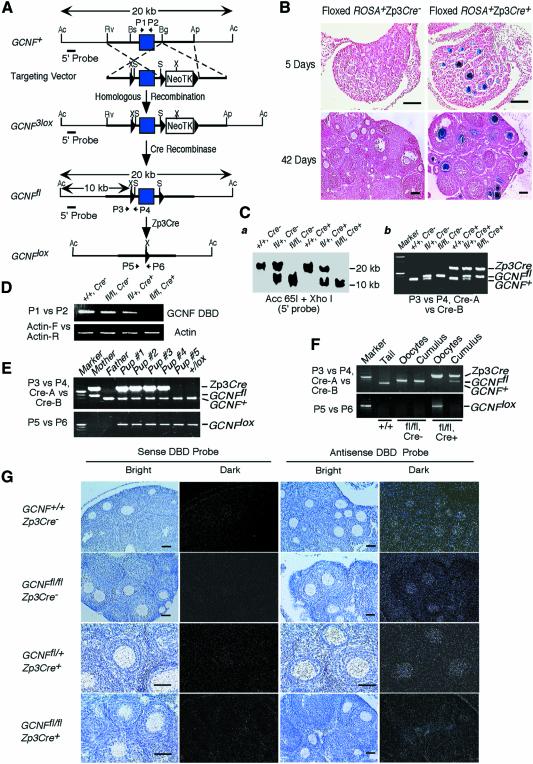
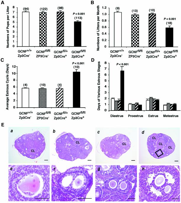
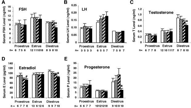
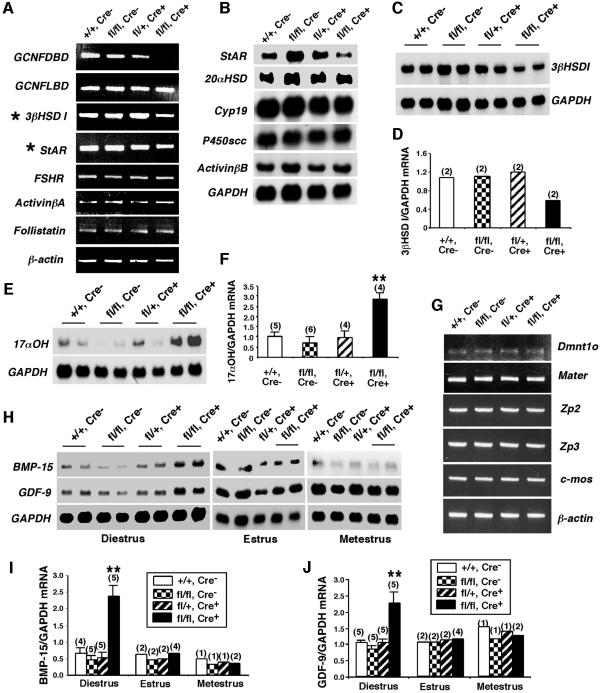
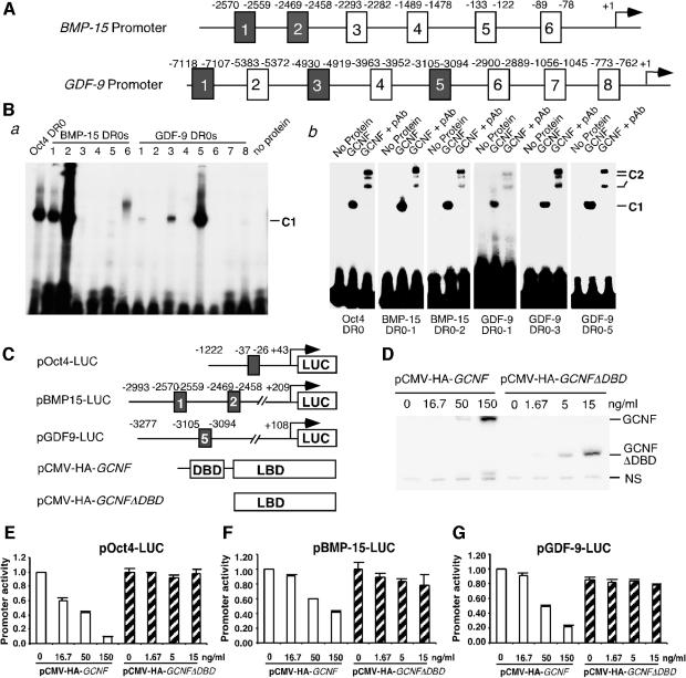
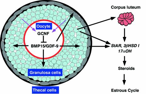
To determine the function of germ cell nuclear factor (GCNF) in female reproduction, we generated an oocyte-specific GCNF knockout mouse model (GCNF(fl/fl)Zp3Cre(+)). These mice displayed hypofertility due to prolonged diestrus phase of the estrous cycle and aberrant steroidogenesis. These reproductive defects were secondary to a primary defect in the oocytes, in which expression of the paracrine transforming growth factor-beta signaling molecules, bone morphogenetic protein 15 (BMP-15) and growth differentiation factor 9 (GDF-9), were up-regulated in GCNF(fl/fl)Zp3Cre(+) females at diestrus. This was a direct effect of GCNF, as molecular studies showed that GCNF bound to DR0 elements within the BMP-15 and GDF-9 gene promoters and repressed their reporter activities. Consistent with these findings, abnormal double-oocyte follicles, indicative of aberrant BMP-15/GDF-9 expression, were observed in GCNF(fl/fl)Zp3Cre(+) females. The Cre/loxP knockout of GCNF in the oocyte has uncovered a new regulatory pathway in ovarian function. Our results show that GCNF directly regulates paracrine communication between the oocyte and somatic cells by regulating the expression of BMP-15 and GDF-9, to affect female fertility.


Figures

None
Fig. 1. Generation of an oocyte-specific GCNF knockout mouse model. (A) Structures of the GCNF3lox, GCNFfl and GCNFlox alleles and the targeting vector. LoxP sites, filled triangles; the DBD-encoding exon, filled box. Restriction enzyme sites of Acc65I (Ac), ApaI (Ap), BglI (Bg), BsiEI (Bs), EcoRV (Rv), XhoI (X) and SalI (S) in each allele are shown. Positions of PCR primers (P1 to P6), and the 5′ probe for Southern blot analysis are indicated. (B) Cre recombinase activity on the floxed ROSA transgene in the ovaries determined by β-galactosidase staining. Bar scale, 200 µm. (C) Genotype analysis of tail biopsies. (a) Southern blot analysis showing a 20 kb band for the GCNF+ allele and a 10 kb band for the GCNFfl allele. (b) PCR analysis using primers (P3 and P4) to distinguish the GCNFfl and GCNF+ alleles, and primers (Cre-A and Cre-B) to determine the presence of Zp3Cre transgene. (D) RT–PCR analysis showing the loss of the DBD encoding region of the GCNF mRNA in the GCNFfl/flZp3Cre+ ovary. (E) Genotyping of tail DNA showing the complete deletion of the GCNF DBD encoding exon in the progenies from the cross between GCNFfl/flZp3Cre+ females and wild-type males. (F) PCR analysis showing complete deletion of the GCNF DBD encoding exon in ovulated oocytes, but not in cumulus cells. (GIn situ hybridization showing the loss of the DBD region of the GCNF mRNA in the oocytes of GCNFfl/flZp3Cre+ mice. Bar scale, 50 µm.
None
Fig. 2. Oocyte-specific GCNF knockout mice display hypofertility, abnormal estrous cycle and double-oocyte follicles. (A) Reduced numbers of pups per litter and (B) reduced numbers of litters per month from the breeding of GCNFfl/flZp3Cre+ females with wild-type males for 1 year. (C) Prolonged length of the estrous cycle in the GCNFfl/flZp3Cre+ females. (D) Prolonged diestrus of the estrous cycle in the GCNFfl/flZp3Cre+ females. (E) Normal ovarian histology except the presence of double oocyte follicles in the GCNFfl/flZp3Cre+ females. (a) GCNF+/+, (b) GCNFfl/fl, (c) GCNFfl/+Zp3Cre+ and (d) GCNFfl/flZp3Cre+ ovaries were 3-month-old littermates. CL, corpus lutea. (e–h) The presence of double-oocyte follicles at the primary (e and f), secondary (g) and antral (h) stages in GCNFfl/flZp3Cre+ ovaries. High magnification of the box in (d) is shown in (h). The bar scale in (a)–(d) is 200 µm. In (e)–(h) the bar scale is 50 µm.
None
Fig. 3. Serum levels of gonadotropins and steroid hormones in the GCNFfl/flZp3Cre+ females during the estrous cycle. (A) FSH, (B) LH, (C) testosterone, (D) estradiol and (E) progesterone levels in the GCNFfl/flZp3Cre+ (filled bar), and three control groups, GCNF+/+ (open bar), GCNFfl/fl (grid bar) and GCNFfl/+Zp3Cre+ (striped bar), are presented as means ± SEs among the indicated numbers of samples in each group in the bar graph. *P < 0.05, when compared with three control groups at diestrus. #P < 0.05, when compared with GCNF+/+ or GCNFfl/fl at estrus.
None
Fig. 4. Expression of ovarian marker genes in the GCNFfl/flZp3Cre+ ovary. (A–F) Expression of key ovarian genes expressed in somatic cells of GCNF+/+ (+/+, Cre–), GCNFfl/fl (fl/fl, Cre–), GCNFfl/+Zp3Cre+ (fl/+, Cre+) and GCNFfl/flZp3Cre+ (fl/fl, Cre+) ovaries at diestrus (day 1–2). (A) RT–PCR showing the reduced StAR and 3βHSD I expression in the GCNFfl/flZp3Cre+ mice. (B–F) Nothern blot analyses showing the mis-expression of (BStAR, (C and D3βHSD I and (E and F17αOH in GCNFfl/flZp3Cre+ mice. Quantitative 3βHSD I and 17αOH mRNA levels in the northern blots are shown in the bar graph in (D) and (F), respectively. (GJ) Expression of oocyte marker genes. (G) RT–PCR showing expression of oocyte genes, Dmnt1o, Mater, Zp2, Zp3 and c-mos, in ovaries. Experiments were repeated twice using two individual animals. (H) Representative radiographs of northern blot analyses showing the BMP-15 and GDF-9 expression. (I and J) Quantitative ovarian (I) BMP-15 and (J) GDF-9 mRNA levels in northern blots. For (F), (I) and (J), relative mRNA levels (normalized to GAPDH mRNA levels) are presented as means ± SEs from various numbers of animal samples (indicated by alphabetic numbers) of each genotype. **P < 0.01 compared with three other groups at diestrus.
None
Fig. 5. Direct regulation of the BMP-15 and GDF-9 expression by GCNF. (A) Schematic representation of putative DR0s in the mouse BMP-15 and GDF-9 promoters. (B) EMSAs showing the binding of in vitro translated GCNF to multiple DR0 elements in the BMP-15 and GDF-9 promoters. (a) GCNF was incubated with different 32P-labeled oligonucleotide probes containing the putative DR0 elements. (b) Retarded DNA-protein complexes in EMSAs by anti-GCNF antibodies (pAb). C1, GCNF–DR0 DNA complexes; C2, antibody retarded GCNF–DR0 DNA complexes. (C) Schematic representation of GCNF expression plasmids and luciferase reporter plasmids containing Oct4, BMP-15 or GDF-9 promoters. (D) Western blot analysis showing the expression levels of full-length GCNF protein and GCNF mutant protein (GCNFΔDBD) in transfected CHO cells using anti-HA antibodies. NS, non-specific protein. (E, G) Dose-dependent repression of the (EOct4, (FBMP-15 and (GGDF-9 expression by GCNF, not GCNFΔDBD, in CHO cells. Promoter activities are presented as means ± SDs of the percentages of the total promoter activities (without pCMV-HA-GCNF plasmid) and represent three independent measurements.
None
Fig. 6. A model for GCNF regulation of BMP-15/GDF-9 signaling during female reproduction. GCNF represses BMP-15/GDF-9 expression, which in turn affects steroidogenesis and female fertility.

Similar articles

Cited by

References

    1. Agoulnik I.Y., Cho,Y., Niederberger,C., Kieback,D.G. and Cooney,A.J. (1998) Cloning, expression analysis and chromosomal localization of the human nuclear receptor gene GCNF. FEBS Lett., 424, 73–78. - PubMed
    1. Bain P.A., Yoo,M., Clarke,T., Hammond,S.H. and Payne,A.H. (1991) Multiple forms of mouse 3 beta-hydroxysteroid dehydrogenase/delta 5-delta 4 isomerase and differential expression in gonads, adrenal glands, liver, and kidneys of both sexes. Proc. Natl Acad. Sci. USA, 88, 8870–8874. - PMC - PubMed
    1. Braat A.K., Zandbergen,M.A., De Vries,E., Van Der Burg,B., Bogerd,J. and Goos,H.J. (1999) Cloning and expression of the zebrafish germ cell nuclear factor. Mol. Reprod. Dev., 53, 369–375. - PubMed
    1. Chen F., Cooney,A.J., Wang,Y., Law,S.W. and O’Malley,B.W. (1994) Cloning of a novel orphan receptor (GCNF) expressed during germ cell development. Mol. Endocrinol., 8, 1434–1444. - PubMed
    1. Chung A.C., Katz,D., Pereira,F.A., Jackson,K.J., DeMayo,F.J., Cooney,A.J. and O’Malley,B.W. (2001) Loss of orphan receptor germ cell nuclear factor function results in ectopic development of the tail bud and a novel posterior truncation. Mol. Cell. Biol., 21, 663–677. - PMC - PubMed

Publication types

MeSH terms

Substances