. 2003 Aug 15;22(16):4132-42.
doi: 10.1093/emboj/cdg414.

Interdimer processing mechanism of procaspase-8 activation

Affiliations

Interdimer processing mechanism of procaspase-8 activation

David W Chang et al. EMBO J. .

Abstract

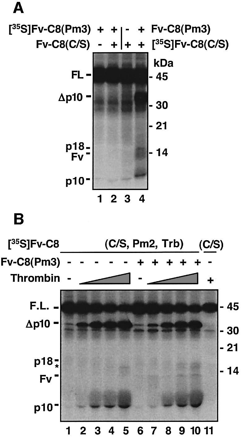
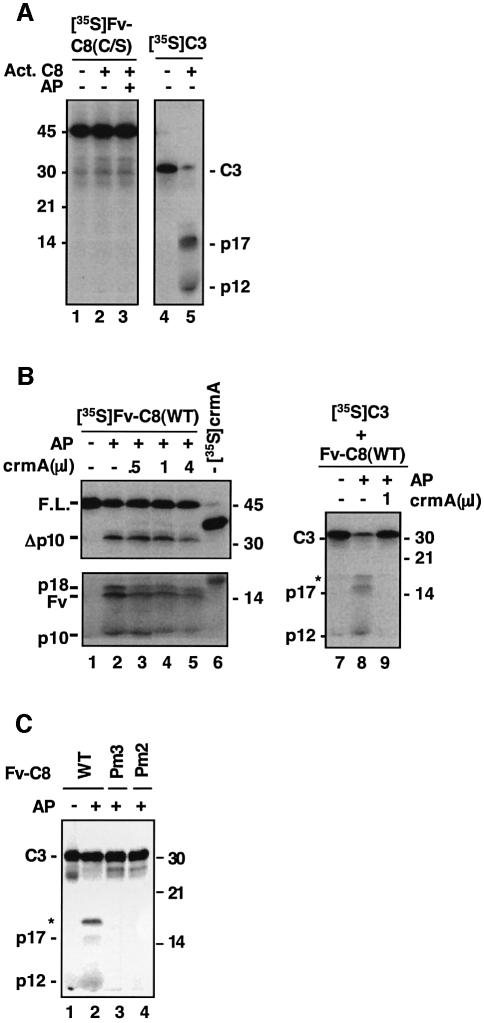
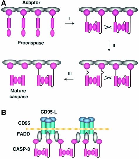
The execution of apoptosis depends on the hierarchical activation of caspases. The initiator procaspases become autoproteolytically activated through a less understood process that is triggered by oligomerization. Procaspase-8, an initiator caspase recruited to death receptors, is activated through two cleavage events that proceed in a defined order to generate the large and small subunits of the mature protease. Here we show that dimerization of procaspase-8 produces enzymatically competent precursors through the stable homophilic interaction of the procaspase-8 protease domain. These dimers are also more susceptible to processing than individual procaspase-8 molecules, which leads to their cross-cleavage. The order of the two interdimer cleavage events is maintained by a sequential accessibility mechanism: the separation of the large and small subunits renders the region between the large subunit and prodomain susceptible to further cleavage. In addition, the activation process involves an alteration in the enzymatic properties of caspase-8; while procaspase-8 molecules specifically process one another, mature caspases only cleave effector caspases. These results reveal the key steps leading to the activation of procaspase-8 by oligomerization.


Figures

None
Fig. 1. Oligomerization-induced caspase-8 activation in vitro. (A) Schematic representation of procaspase-8 and its fusions/mutations: C8(FL), full-length caspase-8; DED, death effector domain; p18 and p10, the large and small subunits that form mature caspase-8; D, cleave site aspartic acid residue; Fv-C8(FL), Fv fusion of full-length caspase-8; Fv, an FK506 binding protein 12 (FKBP12) derivative with the Phe36 to Val mutation; C8(PD), caspase-8 protease domain (amino acids 206–479); Fv-C8, Fv fusion of caspase-8 protein domain; Fv-C8Δ, Fv-C8 lacking amino acids 370–434. Shown on the bottom are various point mutations used in this work: Pm, processing mutant; Im, interface mutant. (B) Inducible activation of caspase-8 in vitro. In vitro translated 35S-labeled Fv-C8 proteins were treated with the indicated concentration of AP20187 (AP). WT, wild type. Reaction mixes were resolved by SDS–PAGE and analyzed by autoradiography. The deduced domain structures of the indicated bands are shown on the left. Arrowheads indicate cleavage sites and the numbers in parentheses mark the order of cleavage. Molecular weight standards are shown on the right. (C) Processing of caspase-8 in the CD95 death-inducing signaling complex (DISC). 35S-labeled full-length wild-type or mutant caspase-8 proteins were treated with CD95 complex isolated from SKW6.4 cells that were either unstimulated (–) or stimulated with an agnostic antibody anti-CD95 (+). Deduced domain structures and molecular weight standards are shown. (D) Effects of catalytic mutants on the processing of wild-type caspase-8. Left and middle panels: 1 µl of in vitro translated [35S]Fv-C8(WT) was treated with AP20187 for 4 h (lanes 1–6) or 2 h (lanes 7–10). In vitro translated non-radiolabeled Fv fusion proteins were added as indicated. Right panel, 1 µl of in vitro translated 35S-labeled proteins was analyzed by autoradiography to show the relative levels of protein expression (lanes 11–14).
None
Fig. 2. Dimerization-induced caspase activity in caspase-8 zymogen. (A) Recombinant Fv-C8 fusions. Purified Fv-C8 proteins (10 µl each) were resolved by SDS–PAGE and visualized by Coomassie staining. Fv-C8(WT) was partially processed during bacterial expression. The effects of dimerization on the protease activity of non-processible and interface caspase-8 mutants are shown in (B) and (C). (B) Fv-C8 proteins (50 ng each) were treated with AP20187 for 4 h and then tested for proteolysis of the fluorogenic substrate IETD-AFC. Lane 14 also contained 10 ng of Fv-C8(C/S). (C) Fv-C8 proteins (50 ng) were incubated with AP20187 (+) or vehicle (–) before being labeled with biotinylated VAD-fmk. Lane 4 contained 10 ng of Fv-C8(C/S). Reaction mixes were separated by SDS–PAGE and analyzed by immunoblotting with either avidin–HRP to detect the labeled species (top) or anti-His6 to detect proteins (bottom). (D) Procaspase-8 in the DISC complex is enzymatically active. CD95 complex from BJAB cells that were either stimulated (+) with anti-CD95 or unstimulated (–). Cell lysates (lys) and the DISC complex were treated with vehicle (–) or with 30 µM (+) or 100 µM (++) biotinylated VAD-fmk. Non-biotinylated z-VAD-fmk was included in the reaction as indicated. The reaction mixes were analyzed by immunoblotting with either avidin–HRP (top) or anti-caspase-8 C15 (bottom). The cell death activity of the caspase-8 precursor is shown in (E) and (F). HeLa cells were transfected with (E) 100 ng of plasmids expressing the indicated Fv-C8 proteins plus an EGFP expressing plasmid and treated with AP20187 (+) or vehicle (–), or (F) 1 µg of plasmids expressing the full-length caspase-8 proteins plus the EGFP plasmid. Apoptosis was scored among GFP-positive cells 18–24 h later.
None
Fig. 3. Procaspase-8 activation requires stable association of the protease domain. (A) Crystal structure of the mature caspase-8 heterotetramer bound to the DEVD-CHO inhibitor (Blanchard et al., 2000) (courtesy of H.Blanchard et al.). Positions of Lys367, Asp395, Ala397 and Lys473, the N-terminus of p10 (Np10) and the C-terminus of p20 (Cp20) are indicated on one half of the tetrameric molecule. (B) Interface mutations that impair the homophilic interaction of the caspase-8 protease domain. Top and middle panels: 35S-labeled caspase-8 protease domains (each contains an HA tag) were mixed with non-radiolabeled protease domains (each contains a FLAG tag), as indicated. The mixes were immunoprecipitated with anti-FLAG antibody M2 conjugated on agarose beads, and the bound proteins (top) and input proteins (middle) were analyzed by autoradiography. Bottom panel: in a parallel experiment, the immunoprecipitation was performed with non-radiolabeled HA-tagged proteins and 35S-labeled FLAG-tagged proteins. The bound FLAG tagged proteins were analyzed by autoradiography. The ImB mutant migrated faster than the other proteins on SDS–PAGE, probably caused by the change of a negatively charged residue to a positive one. (C) Defective processing of the caspase-8 interface mutants. the experiment was performed as in Figure 1B. (D) Full-length interface mutants failed to restore apoptosis in caspase-8-deficient cells. Caspase-8-deficient Jurkat cells were transfected with vector (–) or with constructs expressing either wild-type or mutant full-length caspase-8 proteins, plus pEGFP. Cells were treated with anti-CD95 and the percentage of apoptosis among GFP-positive cells was determined by annexin V staining. None of the interface mutations affected protein stability (data not shown). (E) Restoration of protease activity of interface mutants by c-FLIPL. [35S]Fv-C8(ImA) (lanes 1–3) or [35S]Fv-FLIP (lanes 4–6) was treated with AP20187 (+) or vehicle (–). An equal amount of non-radiolabeled proteins was included as indicated. Similar results were obtained for Fv-C8(ImB) and Fv-C8(ImC) (data not shown).
None
Fig. 4. Dimerization renders procaspase-8 molecules susceptible to cleavage. (A) Spontaneous activation of the FKBP and Fv fusion of full-length caspase-8. In vitro translated Fv-C8(FL) or Fv-C8(FL, C/S) was incubated at 30°C for the indicated duration before being analyzed by SDS–PAGE and autoradiography. Domain structures are shown on the left. Similar results were obtained for the FKBP fusion of caspase-8. (B) Stronger homophilic interaction of Fv-C8(FL). Left panel: mixes of [35S]Fv-C8 (FL) (lane 1 and 2) or [35S]C8(FL) (lanes 3 and 4) (containing an HA tag) with the corresponding non-radiolabeled proteins (+) (containing a FLAG tag) or control in vitro translation mix (–) were immunoprecipitated with anti-FLAG conjugated on agarose beads, and the bound (top) and input (bottom) proteins were analyzed by SDS–PAGE and autoradiography. Right panel: in a parallel experiment, the immunoprecipitation was carried out with non-radiolabeled HA-tagged proteins and 35S-labeled FLAG tagged proteins. (C) Dimerization enhances processing of Fv-C8(C/S) by FKBP-C8(FL). [35S]Fv-C8(C/S) was treated with the indicated non-radiolabeled FKBP fusion proteins in the presence (+) or absence (–) of AP20187. (D) Caspase-8 interface mutations abolished processing of Fv-C8 by FKBP-C8(FL). [35S]Fv-C8(C/S) or Fv-C8(ImA) was treated with FKBP-C8(FL). Similar results were obtained for Fv-C8(ImB) and Fv-C8(ImC) (data not shown). (E) Defective processing of the full-length caspase-8 interface mutants by the DISC. 35S-labeled full-length caspase-8 proteins were treated with the DISC complex as in Figure 1C.
None
Fig. 5. Sequential accessibility mechanism for maintaining the order of cleavage events. (A) A non-processible caspase-8 can process caspase-8(C/S) at both cleavage sites. The indicated combinations of 35S-labeled and non-radiolabeled proteins were treated with AP20187 or vehicle. (B) Severance of the large and small subunits allows for cleavage between the prodomain and the large subunit. [35S]Fv-C8(C/S, Pm2, Trb), which contains the indicated mutations and a thrombin cleavage site inserted between the large and small subunits, or Fv-C8(C/S) was treated with non-radiolabeled Fv-C8(Pm3) in the presence of AP20187. The mixes were incubated for 4 h at 30°C with increasing amounts of thrombin (0.36 × 10–4, 1.2 × 10–4, 3.6 × 10–4 and 12 × 10–4 U/µl), with 12 × 10–4 I/µl thrombin (+) or untreated (–) and analyzed by SDS–PAGE and autoradiography. The size of p18 was slightly larger than the wild-type p18 due to the cleavage at the thrombin site. The asterisk indicates a non-specific cleavage product.
None
Fig. 6. Change of enzymatic characteristics during the activation of procaspase-8. (A) Individual and dimerized caspase-8 precursors cannot be processed by mature caspase-8. [35S]Fv-C8(C/S) or [35S]procaspase-3 (C3) was treated with active caspase-8 (20 ng/µl) and AP20187 as indicated. p17 and p12 are the large and small subunits of mature caspase-3, respectively. (B) The precursor and mature caspase-8 have different substrate specificities. Left panel: lanes 1–5, 1 µl of [35S]Fv-C8(C/S) was treated with AP20187 in the presence of the indicated amount of non-radiolabeled crmA; lane 6 contained 1 µl of 35S-labeled crmA to show the level of protein expression. Right panel: 1 µl of [35S]C3 was treated with Fv-C8 that had been previously treated with AP20187 (+) or with vehicle (–); 1 µl of crmA was included in the reaction as indicated. The asterisk indicates a band containing both the large subunit and the prodomain of caspase-3. (C) Active caspase-8 precursor does not process caspase-3. [35S]C3 was incubated with Fv-C8 proteins that were either pretreated with AP20187 (+) or untreated (–).
None
Fig. 7. The ‘interdimer processing’ model for procaspase-8 activation. (A) Upon adaptor-mediated oligomerization, procaspase–8 molecules in the DISC form dimers through a stable interaction between their protease domains to become proteolytically active (I). These dimers are also susceptible to cleavage by other procaspase-8 dimers at the region linking the large and small subunits (II). The severance of the large and small subunits leads to a conformational change in the region between the prodomain and the large subunit, allowing this region to be subsequently cleaved. The resulting mature caspase-8 is released to the cytosol (III). (B) While two procaspase-8 molecules within a DISC complex form a dimer, the third one may associate with the unpaired caspase-8 from another DISC, allowing the appropriate alignment of these intermediates to facilitate cross-cleavage between the dimers.

Similar articles

Cited by

References

    1. Algeciras-Schimnich A., Shen,L., Barnhart,B.C., Murmann,A.E., Burkhardt,J.K. and Peter,M.E. (2002) Molecular ordering of the initial signaling events of CD95. Mol. Cell. Biol., 22, 207–220. - PMC - PubMed
    1. Ashkenazi A. and Dixit,V.M. (1998) Death receptors: signaling and modulation. Science, 281, 1305–1308. - PubMed
    1. Blanchard H., Donepudi,M., Tschopp,M., Kodandapani,L., Wu,J.C. and Grutter,M.G. (2000) Caspase-8 specificity probed at subsite S(4): crystal structure of the caspase-8-Z-DEVD-CHO complex. J. Mol. Biol., 302, 9–16. - PubMed
    1. Boatright K.M. et al. (2003) A unified model for apical caspase activation. Mol. Cell, 11, 529–541. - PubMed
    1. Chang D.W. and Yang,X. (2003) Activation of procaspases by FK506 binding protein-mediated oligomerization. Sci. STKE, 2003, PL1. - PubMed

Publication types

MeSH terms