. 2003 Aug 15;22(16):4155-65.
doi: 10.1093/emboj/cdg398.

Pak1 and PIX regulate contact inhibition during epithelial wound healing

Affiliations

Pak1 and PIX regulate contact inhibition during epithelial wound healing

Mirjam M P Zegers et al. EMBO J. .

Abstract

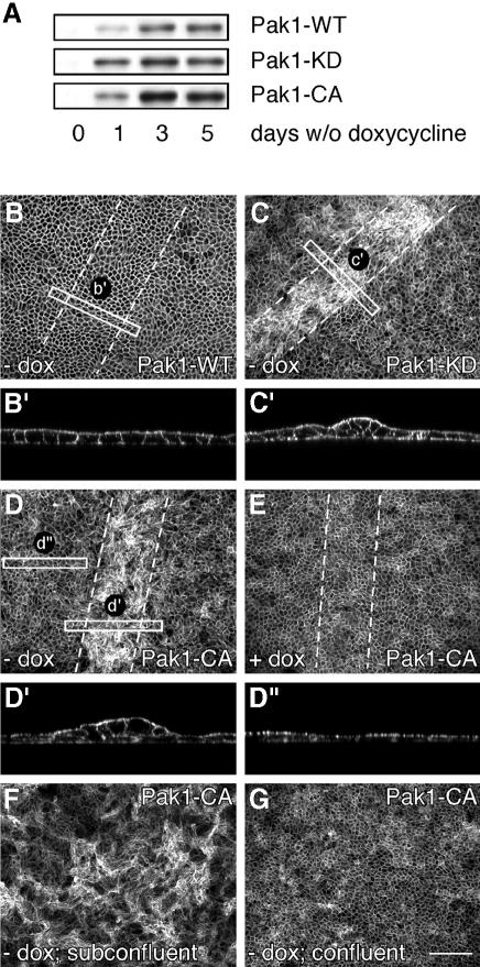
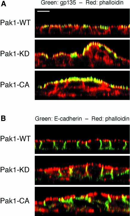
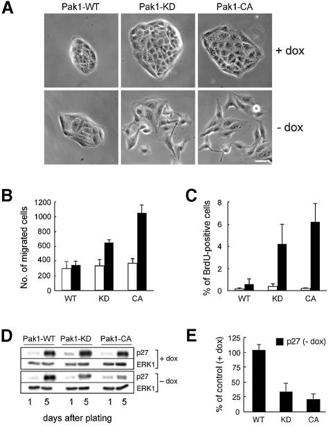
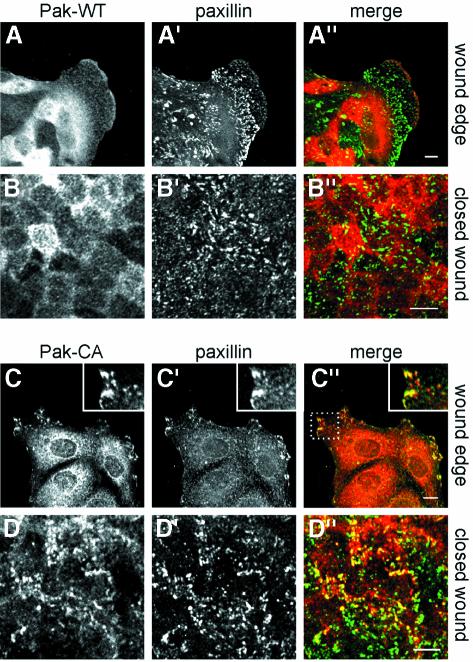
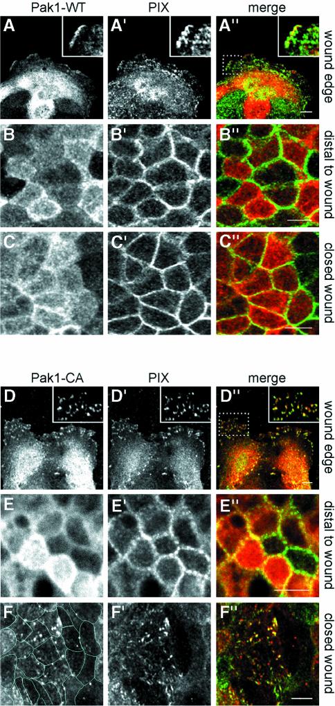
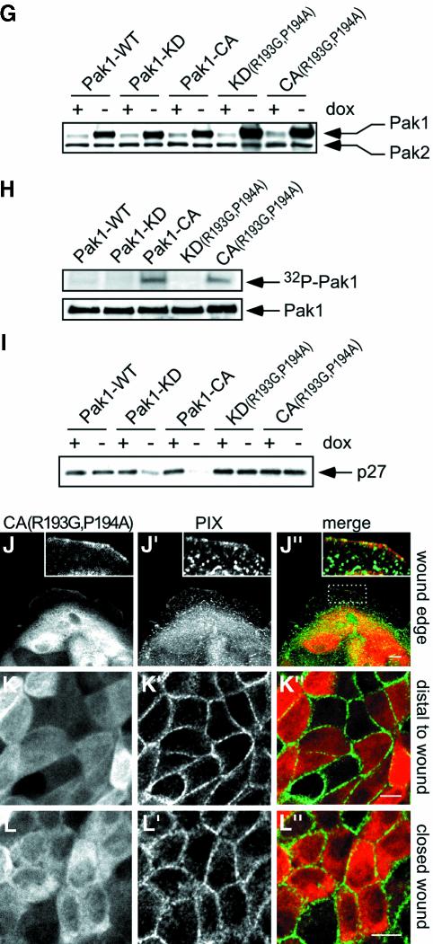
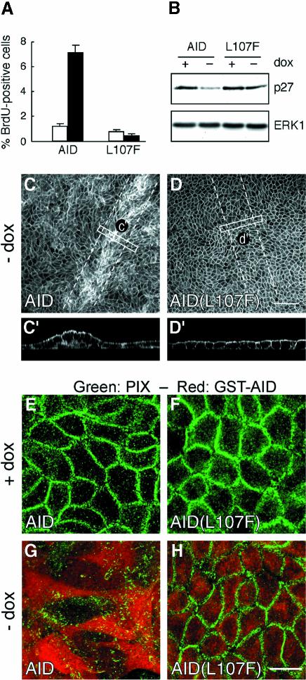
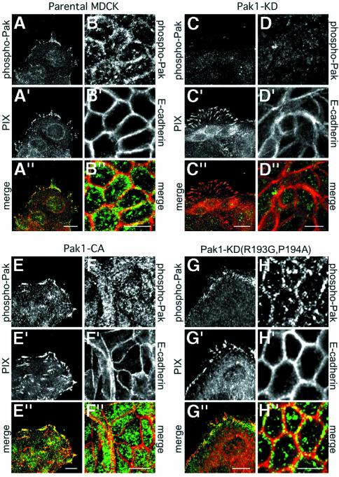
Wound healing in epithelia requires coordinated cell migration and proliferation regulated by signaling mechanisms that are poorly understood. Here we show that epithelial cells expressing constitutively active or kinase-dead mutants of the Rac/Cdc42 effector Pak1 fail to undergo growth arrest upon wound closure. Strikingly, this phenotype is only observed when the Pak1 kinase mutants are expressed in cells possessing a free lateral surface, i.e. one that is not engaged in contact with neighboring cells. The Pak1 kinase mutants perturb contact inhibition by a mechanism that depends on the Pak-interacting Rac-GEF PIX. In control cells, endogenous activated Pak and PIX translocate from focal complexes to cell-cell contacts during wound closure. This process is abrogated in cells expressing Pak1 kinase mutants. In contrast, Pak1 mutants rendered defective in PIX binding do not impede translocation of activated Pak and PIX, and exhibit normal wound healing. Thus, recruitment of activated Pak and PIX to cell-cell contacts is pivotal to transduction of growth-inhibitory signals from neighboring cells in epithelial wound healing.


Figures

None
Fig. 1. Expression of Pak1 mutants interferes with epithelial wound healing. (A) MDCK cells expressing human Pak1-WT, Pak1-KD and Pak1-CA were grown for the indicated periods of time with or without doxycycline (dox). Pak1 expression in cell lysates was assayed by western blotting using an anti-Pak1 antibody, which does not recognize canine Pak1. (B–E) MDCK cells expressing Pak1-WT (B), Pak1-KD (C) or Pak1-CA (D and E) were plated on filters and grown for 5 days in the presence of dox. The monolayers were then wounded by scraping away 30–50 rows of cells (dashed lines indicate wounded areas) and grown for 48 h without (B–D) or with (E) dox. Samples were fixed and stained for F-actin. In non-induced (E) and Pak1-WT-expressing cells (B and enlargement B′) the monolayer restores completely, while wounded areas of Pak1-KD- (C) and Pak1-CA-expressing cells (D) are characterized by cell multilayering (enlargements C′ and D′). Multilayering does not occur further back from the wound edge (enlargement D′′). (F) MDCK cells induced to express Pak1-CA before plating exhibit extensive cell multilayering throughout the culture, whereas no multilayering is observed when Pak1-CA expression is induced after a confluent monolayer is formed (G). Scale bar, 100 µm.
None
Fig. 2. Cells expressing Pak1 kinase mutants retain the capability to polarize. MDCK cells induced to express Pak1-WT, Pak1-KD and Pak1-CA before plating and grown to confluency were stained with phalloidin (red) to outline cell borders and immunolabeled (green) to detect either the apical gp135 marker (A) or E-cadherin (B). Note that, in spite of extensive multilayering, Pak1-KD- and Pak1-CA-expressing cells are still able to segregate gp135 to the free apical surface (A) and E-cadherin to membranes engaged in cell–cell contact (B). Scale bar, 10 µm.
None
Fig. 3. Pak1 mutants promote cell scattering and migration towards HGF and perturb contact inhibition of proliferation. (A) MDCK cells were plated on culture dishes and induced to express Pak1-WT, Pak1-KD or Pak1-CA for 3 days. The images shown are representative of >99% of Pak1-WT-expressing cells, and of 70–90% of Pak1-KD- and Pak1-CA-expressing cells. Scale bar, 20 µm. (B) Migration towards HGF in the lower compartment of Transwell filters was determined as described in Materials and methods. White bars, non-induced controls; black bars, Pak1-expressing cells. In (C) and (D), cells were plated on filters and grown with or without dox. (C) Five days after plating, cell proliferation was assayed by BrdU incorporation as described in Materials and methods. The data represent mean ± SD of three independent experiments. White bars, non-induced controls; black bars, Pak1-expressing cells. (D and E) p27kip1 levels in cell lysates prepared from cells 1 and 5 days after plating were determined by western blotting using a monoclonal anti-p27kip1 antibody (D), and quantified by densitometry (E).
None
Fig. 4. Pak1 mutants fail to redistribute from focal complexes after wound closure. The localization of Pak1 and paxillin in MDCK cells was analyzed before and after wound closure. Images show staining for Pak1 (A–D), paxillin (A′–D′) and merged images (A′′–D′′: Pak1 in red; paxillin in green). In all samples, a diffusely stained intracellular pool of Pak1 is observed. At the wound edge, WT-Pak1 is also found in cell ruffles (A), where limited co-localization with paxillin is observed (A′ and A′′). In contrast, Pak1-CA (C) extensively co-localizes with paxillin (C′ and C′′), although the number of paxillin-containing focal complexes is diminished. Co-localization of Pak1-CA and paxillin is highlighted in the insert in (C′′), which represents an enlargement of the area in the dashed rectangle. In closed wounds, Pak1-WT (B) does not co-localize with paxillin (B′), whereas Pak1-CA (D) remains co-localized with paxillin (D′ and D′′). Pak1-expressing cells comprised ∼80% of all cells, as determined by immunofluoresence. The extent of co-localization of Pak1 and paxillin in Pak1-expressing cells was similar in all Pak1-expressing cells at the indicated sites. Scale bars, 10 µm.
None
Fig. 5. Pak1 regulates PIX localization during epithelial wound closure. Pak1 and PIX localization in MDCK cells at wound edges (AA′′ and DD′′) and within closed wounds (CC′′ and FF′′). In addition, cells >200 µm (∼20 cells) distal to the wound edge were analyzed (BB′′ and EE′′). Images represent staining for Pak1 (A–F), PIX (A′–F′) and Pak1 (red) and PIX (green) merged (A′′–F′′). Inserts in (A′′) and (D′′) show co-localization of Pak1 and PIX and represent enlargements of areas enclosed by rectangles. (A–C) Pak1-WT-expressing cells. At wound edges, Pak1-WT accumulates in cell ruffles (A), and shows limited co-localization with PIX (A′). In cells distal to wound edges and in closed wounds, Pak1-WT partly accumulates at sites of cell–cell contacts (B and C) and co-localizes with PIX (B′ and C′), which is concentrated at areas of cell–cell contact. (D–F) Pak1-CA-expressing cells. Pak1-CA (D) and PIX (D′) co-localize extensively in focal contacts in cells at the wound edge (D–D′′, and insert in D′′). In cells distal to the wound edge, Pak1-CA (E) localizes to areas of cell–cell contact and diffusely within the cytoplasm, while PIX (E′) is concentrated at cell–cell contacts. In closed wounds, Pak1-CA (F) and PIX (F′) co-localize in focal contact-like structures while PIX staining at cell–cell contacts is largely absent. Lines in (F) outline individual cells. (G–I) Cell lysates were prepared from cells grown for 5 days with or without dox. (G) Pak expression was analyzed by probing western blots with an antibody, which recognizes Pak1 and Pak2 in MDCK cells. In the absence of dox, Pak1-WT, Pak1-KD and Pak1-CA overexpression levels are 3- to 4-fold, whereas Pak1-KD(R193G,P194A) and Pak-CA(R193G,P194A) are overexpressed ∼8-fold. (H) Kinase activity of Pak1 mutants was determined by autophosphorylation (top panel) of Pak immunoprecipitates (bottom panel) as described in Materials and methods. (I) Western blot showing levels of p27kip1. (J–L) Pak1-CA(R193G,P194A)-expressing cells. In these cells, the localization of Pak1 (JL) and PIX (J′–L′) and their merged distribution (J′′–L′′) is virtually identical to those observed with cells expressing Pak1-WT. Pak1-expressing cells comprised 70–80% of all cells. Images are representative for all Pak1-expressing cells at the indicated sites. Scale bars, 10 µm.
None
Fig. 6. Pak1 AID perturbs PIX redistribution and contact inhibition during wound healing. MDCK cells expressing GST-tagged Pak1 AID or the inactive Pak1 AID(L107F) were grown for 5 days with or without dox. (A) Cell proliferation analyzed by BrdU incorporation. (B) Western blot showing p27 levels. (C and D) Five-day-old monolayers of filter grown MDCK cells were scrape wounded as indicated, and induced for 48 h, resulting in cell multilayering in closed wounds of AID- (C) but not in AID(L107F)-expressing (D) cells. (E–H) The localization of PIX and AID (E and G) or AID(L107F) (F and H) in closed wounds was determined by immunostaining with monoclonal anti-PIX (green) and polyclonal anti-GST (red) antibodies, respectively. Expression of AID (G), but not AID(L107F) (H) prevents the relocalization of PIX to cell–cell contacts after wound closure. AID- and AID(L107F)-expressing cells comprised 80–90% of all cells. Images are representative for all AID/AID(L107F)-expressing cells. Scale bar in (C and D), 100 µm; (E–H), 10 µm.
None
Fig. 7. Pak1 kinase mutants perturb recruitment of endogenous Pak to lateral membranes. The localization of activated, phosphorylated Pak (phospho-Pak) was analyzed in cells at wound edges and within closed wounds. Staining of untransfected, T23 MDCK cells reveals that endogenous phospho-Pak is present in focal complexes in cells at wound edges (A) where it co-localizes with PIX (A′ and A′′). After wound closure, endogenous phospho-Pak relocalizes to areas of cell–cell contacts (B), which are delineated by staining with E-cadherin (B′ and B′′). Cells expressing Pak1-KD (∼80% of all cells) exhibit a virtual absence of phospho-Pak staining at wound edges (CC′′) as well as at cell–cell contacts (DD′′). In Pak1-CA-expressing cells (∼85% of all cells), phospho-Pak staining is increased at focal contacts (EE′′), but is absent from cell–cell contacts (FF′′). Localization of phospho-Pak in cells expressing Pak1-KD(P193G,R194A) is virtually similar to untransfected control cells, both at wound edges (GG′′) and at cell–cell contacts in closed wounds (HH′′). Images are representative for all Pak1-expressing cells. Scale bars, 10 µm.

Similar articles

Cited by

References

    1. Bagrodia S. and Cerione,R.A. (1999) Pak to the future. Trends Cell Biol., 9, 350–355. - PubMed
    1. Bagrodia S., Taylor,S.J., Jordon,K.A., Van Aelst,L. and Cerione,R.A. (1998) A novel regulator of p21-activated kinases. J. Biol. Chem., 273, 23633–23636. - PubMed
    1. Bagrodia S., Bailey,D., Lenard,Z., Hart,M., Guan,J.L., Premont,R.T., Taylor,S.J. and Cerione,R.A. (1999) A tyrosine-phosphorylated protein that binds to an important regulatory region on the cool family of p21-activated kinase-binding proteins. J. Biol. Chem., 274, 22393–22400. - PubMed
    1. Brown M.C., West,K.A. and Turner,C.E. (2002) Paxillin-dependent paxillin kinase linker and p21-activated kinase localization to focal adhesions involves a multistep activation pathway. Mol. Biol. Cell, 13, 1550–1565. - PMC - PubMed
    1. Daniels R.H., Hall,P.S. and Bokoch,G.M. (1998) Membrane targeting of p21-activated kinase 1 (PAK1) induces neurite outgrowth from PC12 cells. EMBO J., 17, 754–764. - PMC - PubMed

Publication types

MeSH terms

Substances