. 2003 Aug 15;22(16):4223-36.
doi: 10.1093/emboj/cdg397.

Regulation of PI4,5P2 synthesis by nuclear-cytoplasmic shuttling of the Mss4 lipid kinase

Affiliations

Regulation of PI4,5P2 synthesis by nuclear-cytoplasmic shuttling of the Mss4 lipid kinase

Anjon Audhya et al. EMBO J. .

Abstract

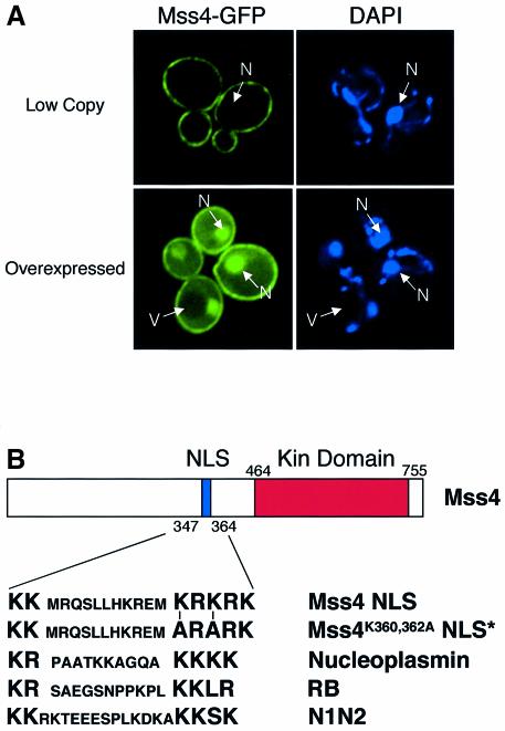
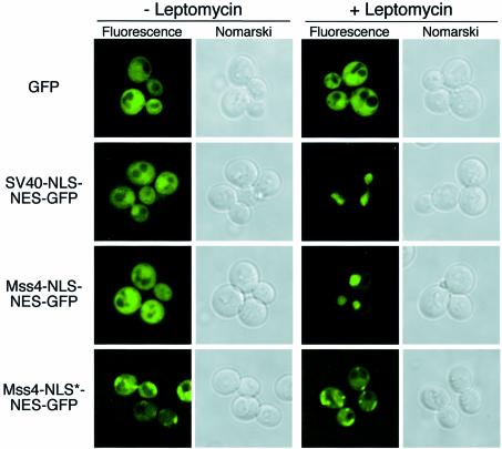
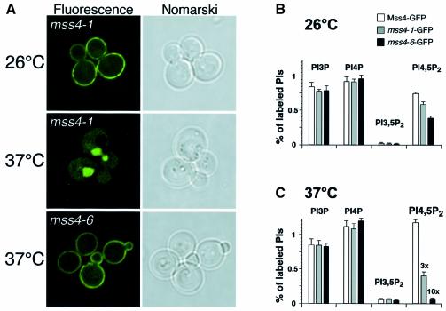
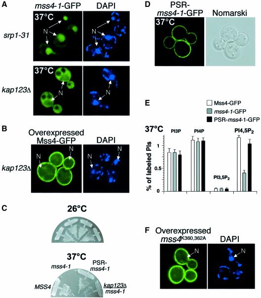
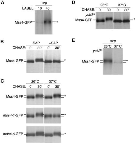
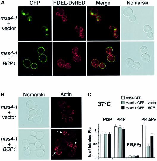
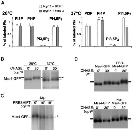
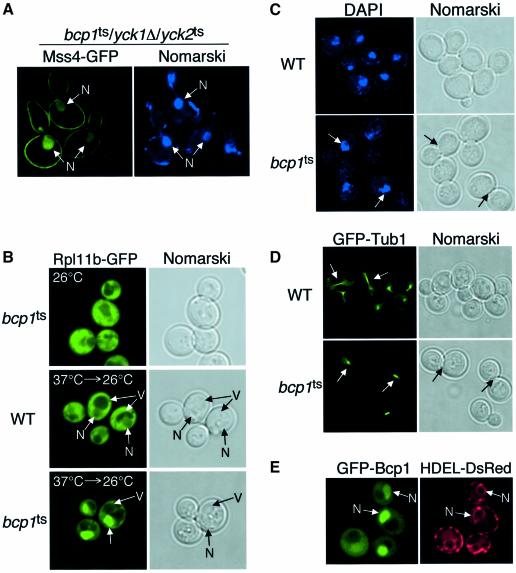
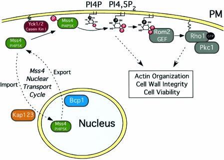
The essential phospholipid PI4,5P(2) is generated by a well conserved PI4P 5-kinase, Mss4, in yeast. Balanced production and turnover of PI4,5P(2) is important for normal organization of the actin cytoskeleton and cell viability. Previous studies have shown that multiple PI phosphatases can regulate PI4,5P(2) levels. We report a new, unexpected regulatory mechanism for PI4,5P(2) homeostasis, directed by nuclear-cytoplasmic shuttling of the lipid kinase. We show that Mss4 is a phosphoprotein, which contains a functional nuclear localization signal (NLS) and can shuttle between the cytoplasm and the nucleus. Temperature-conditional mss4 cells that accumulate Mss4 protein in the nucleus exhibit reduced levels of PI4,5P(2), depolarization of the actin cytoskeleton and a block in Mss4 phosphorylation, suggesting an essential role for phosphorylated Mss4 at the plasma membrane. Through the isolation of gene dosage-dependent suppressors of mss4 mutants, we identified Bcp1, a protein enriched in the nucleus, which is required for Mss4 nuclear export and is related to the mammalian BRCA2-interacting protein BCCIP. Together, these studies suggest a new mechanism for lipid kinase regulation through regulated nuclear-cytoplasmic shuttling.


Figures

None
Fig. 1. Mss4 is localized to the plasma membrane and the nucleus. (Amss4Δ cells carrying pRS416 MSS4–GFP (Low Copy) or pRS426 MSS4–GFP (Overexpressed) were visualized by fluorescence microscopy. DAPI-stained DNA is shown on the right. Pictures are representative of >150 cells observed. Nuclei (N) and vacuoles (V) are indicated by arrows. (B) Cartoon showing the structure of Mss4, including the sequence of its potential bipartite NLS, a mutated form of the NLS (NLS*), and an alignment with other previously characterized NLSs.
None
Fig. 2. Mss4 harbors a functional nuclear localization signal. crm1Δ cells expressing crm1T539C were transformed with constructs expressing GFP, SV40-NLS–NES–GFP, Mss4-NLS–NES–GFP or Mss4-NLS*–NES–GFP, and visualized by fluorescence microscopy in the presence or absence of 100 ng/ml leptomycin. Pictures are representative of >150 cells observed.
None
Fig. 3. Inappropriate nuclear import of Mss4 results in a defect in PI4,5P2 production. (Amss4Δ cells expressing mss4-1GFP or mss4-6GFP were visualized by fluorescence microscopy at the temperatures indicated. (B and C) Phosphoinositide levels in mss4Δ cells expressing MSS4–GFP, mss4-1–GFP or mss4-6–GFP.
None
Fig. 4. Mss4 plays an essential role in the cytoplasm but not in the nucleus. (Asrp1-31 or kap123Δ cells expressing mss4-1–GFP were visualized by fluorescence microscopy after shift to 37°C. DAPI-stained DNA is shown on the right. Nuclei (N) are indicated by arrows. (Bkap123Δ cells overexpressing Mss4–GFP. (Cmss4Δ or mss4Δ/kap123Δ cells expressing MSS4–GFP, mss4-1–GFP or PSR-mss4-1–GFP were grown at 26 or 37°C for 60 h. (Dmss4Δ cells expressing PSR-mss4-1–GFP were visualized by fluorescence microscopy after shift to 37°C. (E) Phosphoinositide levels in mss4Δ cells expressing MSS4–GFP, mss4-1–GFP or PSR-mss4-1–GFP. (Fmss4Δ cells overexpressing mss4K360A,K362A–GFP were visualized by fluorescence microscopy.
None
Fig. 5. Mss4 is a substrate for casein kinase I at the plasma membrane. (A and Emss4Δ or yck1Δ/yck2ts cells expressing MSS4–GFP were metabolically labeled at the appropriate temperature for the indicated time with 32P-labeled orthophosphate. Extracts were immunoprecipitated with anti-GFP antibodies, followed by SDS–PAGE analysis and autoradiography. (BDmss4Δ cells or yck1Δ/yck2ts expressing MSS4–GFP, mss4-1–GFP or mss4-6–GFP were metabolically labeled for 10 min with 35S-labeled cysteine and methionine, and chased for the indicated time. Extracts were immunoprecipitated with anti-GFP antibodies, incubated in the presence or absence of shrimp alkaline phosphatase, and subjected to SDS–PAGE analysis followed by autoradiography. Asterisks indicate the mobility of hyperphosphorylated Mss4.
None
Fig. 6. Bcp1 regulates Mss4 nuclear export. (Amss4Δ cells expressing mss4-1–GFP or HDEL-DsRed, and those overexpressing either empty vector or a plasmid carrying BCP1, were visualized by fluorescence microscopy. (Bmss4Δ cells expressing mss4-1–GFP and overexpressing either empty vector or a plasmid carrying BCP1 were stained with rhodamine–phalloidin and visualized by fluorescence microscopy. Arrows indicate buds with polarized actin localization. (C) Phosphoinositide levels in mss4Δ cells expressing MSS4–GFP or mss4-1–GFP, and overexpressing vector alone or a plasmid carrying BCP1.
None
Fig. 7. Bcp1 is required for appropriate function and localization of Mss4. (A) Phosphoinositide levels in bcp1Δ cells expressing wild-type BCP1 or bcp1ts. (Bbcp1ts cells expressing MSS4–GFP were treated as described in Figure 5C. (Cbcp1ts cells expressing MSS4–GFP were preincubated at 37°C for the indicated time and metabolically labeled with 32P-labeled orthophosphate. Extracts were treated as described in Figure 5A. Asterisks indicate the mobility of hyperphosphorylated Mss4. (D) Wild-type or bcp1ts cells expressing MSS4–GFP or PSR-MSS4–GFP were metabolically labeled at 37°C for 10 min with 35S-labeled cysteine and methionine, and chased for the indicated time. Extracts were treated as above.
None
Fig. 8. Bcp1 is involved in nuclear protein export. (Abcp1ts/yck1Δ/yck2ts cells expressing Mss4–GFP were visualized using fluorescence microscopy at the indicated temperature. DAPI-stained DNA is shown on the right. Nuclei (N) are indicated by arrows. (B) Wild-type or bcp1ts cells expressing RPL11B–GFP were visualized using fluorescence microscopy at the indicated temperature. Nuclei (N) and vacuoles (V) are indicated by arrows. (C) Wild-type or bcp1ts cells were incubated at 37°C for 2 h, fixed and stained with DAPI, and visualized by fluorescence microscopy. Arrows highlight the location of DAPI-stained nuclei. (D) Wild-type or bcp1ts cells expressing GFP–TUB1 were incubated at 37°C for 2 h and visualized by fluorescence microscopy. Arrows highlight the mitotic spindle. (Ebcp1Δ cells expressing GFP–BCP1 and HDEL-DsRED were visualized by fluorescence microscopy.
None
Fig. 9. Model for the regulation of Mss4 localization/activity involving phosphorylation by Yck1/2, nuclear import by Kap123 and nuclear export by Bcp1. The plasma membrane is denoted by PM.

References

    1. Audhya A. and Emr,S.D. (2002) Stt4 PI 4-kinase localizes to the plasma membrane and functions in the Pkc1-mediated MAP kinase cascade. Dev. Cell, 2, 593–605. - PubMed
    1. Audhya A., Foti,M. and Emr,S.D. (2000) Distinct roles for the yeast phosphatidylinositol 4-kinases, Stt4p and Pik1p, in secretion, cell growth and organelle membrane dynamics. Mol. Biol. Cell, 11, 2673–2689. - PMC - PubMed
    1. Berridge M.J. and Irvine,R.F. (1984) Inositol trisphosphate, a novel second messenger in cellular signal transduction. Nature, 312, 315–321. - PubMed
    1. Bevis B.J., Hammond,A.T., Reinke,C.A. and Glick,B.S. (2002) De novo formation of transitional ER sites and Golgi structures in Pichia pastoris. Nat. Cell Biol., 4, 750–756. - PubMed
    1. Boronenkov I.V., Loijens,J.C., Umeda,M. and Anderson,R.A. (1998) Phosphoinositide signaling pathways in nuclei are associated with nuclear speckles containing pre-mRNA processing factors. Mol. Biol. Cell, 9, 3547–3560. - PMC - PubMed

Publication types

MeSH terms