. 2003 Aug 15;22(16):4294-303.
doi: 10.1093/emboj/cdg404.

Localization of proteins bound to a replication origin of human DNA along the cell cycle

Affiliations

Localization of proteins bound to a replication origin of human DNA along the cell cycle

Gulnara Abdurashidova et al. EMBO J. .

Abstract

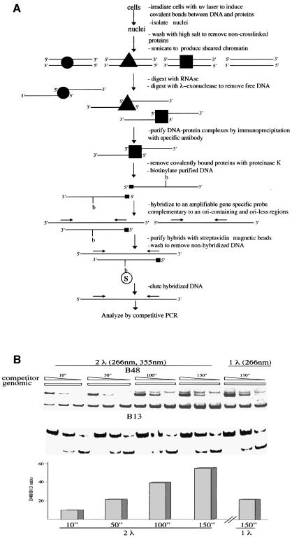
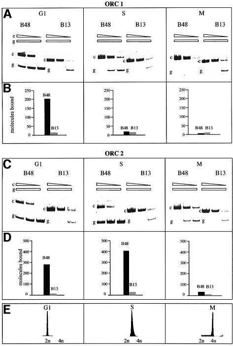
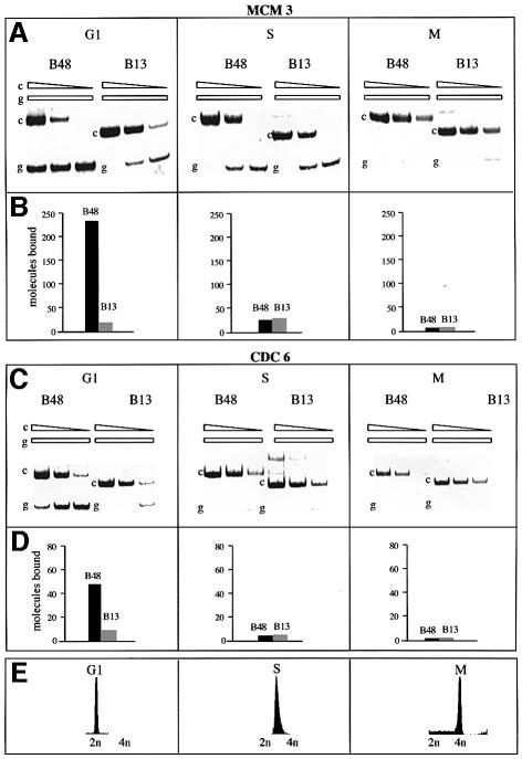
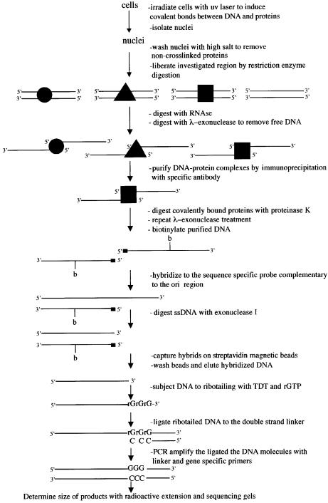
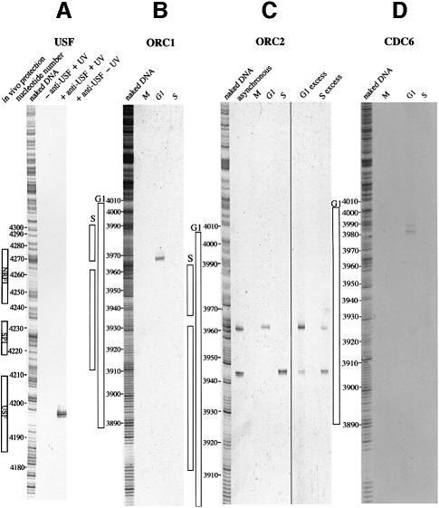
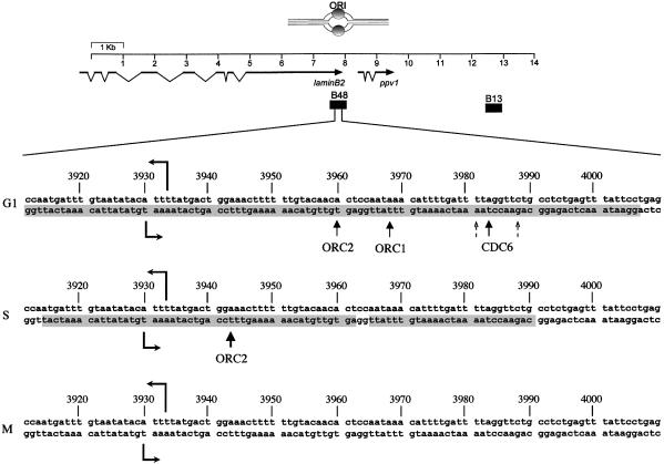
The proteins bound in vivo at the human lamin B2 DNA replication origin and their precise sites of binding were investigated along the cell cycle utilizing two novel procedures based on immunoprecipitation following UV irradiation with a pulsed laser light source. In G(1), the pre-replicative complex contains CDC6, MCM3, ORC1 and ORC2 proteins; of these, the post-replicative complex in S phase contains only ORC2; in M phase none of them are bound. The precise nucleotide of binding was identified for the two ORC and the CDC6 proteins near the start sites for leading-strand synthesis; the transition from the pre- to the post-replicative complex is accompanied by a 17 bp displacement of the ORC2 protein towards the start site.


Figures

None
Fig. 1. Procedure for the identification of proteins bound to the lamin B2 ori sequence and validation with the USF binding. (A) Flow-chart of the procedure; for a detailed description see Materials and methods. (B) Cross-linking of USF to the ori sequence: the cells were exposed to the treatment described in (A) and the isolated DNA was analyzed for the abundance of ori (B48 probe) and non-ori (B13 probe) sequences. From left to right, the results of five sets of competitive PCR experiments (Giacca et al., 1997) are shown: the first four performed on DNA isolated after treatment of the cells with two-wavelength pulsed laser light for the indicated times and the fifth one with one wavelength light and one exposure time only. The immunoprecipitation was performed with anti-USF antibody. Each set of PCR reactions contained the same amount of isolated DNA and concentrations of competitor decreasing by a factor of 10. The upper portion of the figure shows the results of the analysis with a competitor for the ori sequence, the lower portion with a competitor for the non-ori sequence. The amounts of the corresponding sequences in genomic DNA are interpolated by comparison with the known abundance of competitor molecules present. The quantitation of the relative abundance of ori versus non-ori sequence is shown at the bottom. The two-wavelength treatment appears significantly more efficient.
None
Fig. 2. Presence of hOrc1p and hOrc2p on the ori sequence along the cell cycle. The cells arrested in the G1, S or M phase were irradiated with two-wavelength light for 150 s and treated with the procedure described in Figure 1A. The isolated DNA was analyzed for the abundance of ori and non-ori sequences as described in Figure 1B. (A and C) PCR analysis following precipitation with antibodies versus hOrcp1 and hOrcp2, respectively. (B and D) Quantitation of the abundance of ori and non-ori sequences interpolated from the PCR analysis. (E) Flow-cytometric analysis of the treated cell cultures.
None
Fig. 3. Presence of hMcm3p and hCdc6p on the ori sequence along the cell cycle. As Figure 2, except that the antibodies were those raised versus the indicated proteins.
None
Fig. 4. Summary of the presence of DNA replication proteins on the ori sequence along the cell cycle. The data of the experiments reported in Figures 2 and 3 are reported for the four investigated proteins as the relative abundance of ori versus non-ori sequences in the DNA isolated after treatment with the appropriate antibody at different time points of the cell cycle.
None
Fig. 5. Procedure for the identification of the nucleotide(s) bound in vivo by a particular protein. For a detailed description see Materials and methods.
None
Fig. 6. Validation of the procedure with USF and identification of the binding sites of hOrc1p, hOrc2p and hCdc6p on the ori region at different time points of the cell cycle. (A) Asynchronously growing cells were treated with the procedure reported in Figure 5 utilizing anti-USF antibody. The results obtained with non-irradiated and non-immunoprecipitated controls are shown. On the left, the nucleotide position identified from the parallel treatment of naked DNA is shown. The protections observed in vivo (Dimitrova et al., 1996) on the PPV1 gene promoter are also shown. (B–D) Results of the application of the procedure described in Figure 5 to cells in different conditions of synchronization. (B) Anti-hOrc1p antibody; (C) anti-hOrc2p antibody; (D) anti-hCdc6 antibody. The cell cycle phase is indicated at the top of each lane. The nucleotide positions identified from the parallel treatment of naked DNA and the protection observed in vivo in G1 or S on the ori sequence (Abdurashidova et al., 1998) are reported.
None
Fig. 7. Variations in the localization of hOrc1p, hOrc2p and hCdc6p at different time points of the cell cycle. Top, a map of the ori region, indicating the position of the surrounding genes and of the probes utilized for ori (B48) and non-ori DNA. Below, a 100 nucleotide portion of the ori region is shown. The two oppositely directed horizontal arrows indicate the start sites of leading-strand synthesis (Abdurashidova et al., 2000). The shadowed portions of the sequence show the protection observed in vivo on the lower strand in the indicated phase of the cell cycle (Abdurashidova et al., 1998). The upward pointing arrows indicate the nucleotide bound by the three proteins in each phase.

References

    1. Abdurashidova G., Riva,S., Biamonti,G., Giacca,M. and Falaschi,A. (1998) Cell cycle modulation of protein–DNA interactions at a human replication origin. EMBO J., 17, 2961–2969. - PMC - PubMed
    1. Abdurashidova G., Deganuto,M., Klima,R., Riva,S., Biamonti,G., Giacca,M. and Falaschi,A. (2000) Start sites of bidirectional DNA synthesis at the human lamin B2 origin. Science, 287, 2023–2026. - PubMed
    1. Aparicio O.M., Weinstein,D.M. and Bell,S.P. (1997) Components and dynamics of DNA replication complexes in S.cerevisiae: redistribution of MCM proteins and Cdc45p during S phase. Cell, 91, 59–69. - PubMed
    1. Bell S.P. and Stillman,B. (1992) ATP-dependent recognition of eukaryotic origins of DNA replication by a multiprotein complex. Nature, 357, 128–134. - PubMed
    1. Biamonti G. et al. (1992) The gene for a novel human lamin maps at a highly transcribed locus of chromosome 19 which replicates at the onset of S-phase. Mol. Cell. Biol., 12, 3499–3506. - PMC - PubMed

Publication types

MeSH terms