. 2003 Aug 15;22(16):4304-13.
doi: 10.1093/emboj/cdg406.

Elg1 forms an alternative RFC complex important for DNA replication and genome integrity

Affiliations

Elg1 forms an alternative RFC complex important for DNA replication and genome integrity

Mohammed Bellaoui et al. EMBO J. .

Abstract

Genome-wide synthetic genetic interaction screens with mutants in the mus81 and mms4 replication fork-processing genes identified a novel replication factor C (RFC) homolog, Elg1, which forms an alternative RFC complex with Rfc2-5. This complex is distinct from the DNA replication RFC, the DNA damage checkpoint RFC and the sister chromatid cohesion RFC. As expected from its genetic interactions, elg1 mutants are sensitive to DNA damage. Elg1 is redundant with Rad24 in the DNA damage response and contributes to activation of the checkpoint kinase Rad53. We find that elg1 mutants display DNA replication defects and genome instability, including increased recombination and mutation frequencies, and minichromosome maintenance defects. Mutants in elg1 show genetic interactions with pathways required for processing of stalled replication forks, and are defective in recovery from DNA damage during S phase. We propose that Elg1-RFC functions both in normal DNA replication and in the DNA damage response.


Figures
None
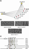
Fig. 1. Genome-wide synthetic lethal screens with mus81Δ and mms4Δ identify the RFC homolog Elg1. (A) The results from synthetic genetic array analysis with mus81Δ and mms4Δ presented as a genetic interaction map. Lines connecting genes represent synthetic lethality or synthetic slow growth. Red circles indicate novel genetic interactions. (B) Tetrad confirmation of the elg1Δ crosses. Each column represents the four spores from a single ascus. Double mutant colonies, as detected by selection for the dominant selectable marker linked to each gene, are indicated by white arrowheads. (C) Schematic representation of the conserved sequence blocks in the S.cerevisiae RFC family genes. Elg1 contains six of the seven RFC boxes found in Rfc1.
None
Fig. 2. Elg1 forms complexes with Rfc2, 3, 4 and 5, but not with Rfc1, Rad24 or Ctf18. (A) Extracts from yeast strains expressing the indicated epitope-tagged RFC proteins were immunoprecipitated with antibody against the flag epitope. Ten percent of the input extract (I) and the immunoprecipitate (P) were fractionated by SDS–PAGE. Immunoblots were probed with anti-flag antibody to detect Rfc1, Rfc2, Rfc3, Rfc4 and Rfc5, and with anti-myc antibody to detect Elg1. A non-specific cross-reacting polypeptide is indicated (*). (B) Extracts from strains expressing the indicated proteins were immunoprecipitated with anti-flag antibody to precipitate Rfc5 and Rad24. Immunoblots were probed with anti-flag antibody to detect Rfc5 and Rad24 and with anti-myc to detect Elg1. (C) Extracts were immunoprecipitated with anti-HA antibody to precipitate Elg1. Immunoblots were probed with anti-flag antibody to detect Rfc5, anti-myc to detect Ctf18 and anti-HA to detect Elg1.
None
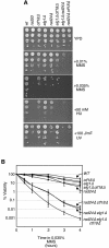
Fig. 3. Elg1 is required for the DNA damage response. (A) Ten-fold serial dilutions of cultures of the indicated mutants were spotted on YPD, YPD containing 0.01% (v/v) MMS, 0.035% (v/v) MMS, 50 mM HU, or on YPD that was subsequently exposed to 100 J/m2 UV. Plates were incubated at 30°C for 2–3 days. (B) Logarithmically growing cultures of the indicated mutants were incubated in YPD containing 0.035% (v/v) MMS at 30°C. At the indicated times, samples were withdrawn and plated on medium lacking MMS to determine the number of viable cells. The percentage of viable cells relative to the number of viable cells at t = 0 is shown. Plots represent the average of three experiments, and error bars span 1 SD.
None
Fig. 4. Rad53 activation defects in elg1Δ. (A) S–M checkpoint. Logarithmically growing cultures were arrested in G1 with α-factor and released into medium containing 200 mM HU. At the indicated times, samples were fixed and extracts fractionated by SDS–PAGE. Following transfer, the immunoblot was probed with anti-Rad53 antibody. Phosphorylation of Rad53 causes a shift in electrophoretic mobility (Rad53-P) and is a marker for checkpoint activation. (B) Intra-S phase checkpoint. Logarithmically growing cultures were treated with 0.035% (v/v) MMS. At the indicated times, samples were withdrawn and Rad53 activation was analyzed by immunoblotting. (C) Cell cycle progression in the presence of MMS was assessed by flow cytometry. Logarithmically growing cultures were treated with 0.035% (v/v) MMS. At the indicated times, samples were fixed and the DNA contents of cells in each sample were analyzed by flow cytometry. The positions of cells with 1C and 2C DNA contents are indicated on the histograms.
None
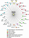
Fig. 5. Genome-wide synthetic genetic screens with elg1Δ identify homologous recombination, fork re-start and S phase checkpoint pathways. The results of synthetic genetic array analysis with elg1Δ presented as a genetic interaction map. Lines connecting genes represent synthetic lethality or synthetic slow growth. Colored circles designate the cellular role of the interacting genes.
None
Fig. 6. elg1Δ mutants display DNA replication defects. (A) Progression through S phase. Wild-type or elg1Δ cells were arrested in G1 (t = 0) and released synchronously into the cell cycle. Samples were removed at the indicated times and analyzed by flow cytometry. The shaded histograms represent the cell cycle distribution of the asynchronous cultures before the G1 arrest. Overlaid histograms represent the cell cycle distribution at the indicated times after release from the G1 arrest. The positions of cells with 1C and 2C DNA contents are indicated. (B) Plasmid loss in wild-type, elg1Δ and ctf19Δ. The probability of plasmid loss per generation is plotted, and error bars span 1 SD. (C) Suppression of elg1Δ MMS sensitivity by PCNA overexpression. Serial dilutions of wild-type or elg1Δ cells carrying empty vector (v) or GAL1-POL30 plasmid (POL30) were plated on synthetic medium with 2% glucose (Glu; uninduced) or 2% galactose + 2% raffinose (Gal; induced), plus or minus 0.01% MMS.
None
Fig. 7. elg1Δ mutants are defective in recovery from MMS-induced replication fork stalling. (A) S phase progression in the presence of MMS. Wild-type or elg1Δ cells were arrested in G1 (t = 0) and released synchronously into medium containing 0.035% (v/v) MMS. Samples were removed at the indicated times and analyzed by flow cytometry. The shaded histograms represent the cell cycle distribution of the asynchronous cultures before the G1 arrest. Overlaid histograms represent the cell cycle distribution at the indicated times after release from the G1 arrest. (B) Checkpoint activation of Rad53 during recovery from MMS damage. Cells were arrested in G1, released into MMS for 1 h, and then transferred to medium lacking MMS (t = 0). At the indicated times, samples were withdrawn and Rad53 activation was analyzed by immunoblotting.

Similar articles

Cited by

References

    1. Aguilera A., Chavez,S. and Malagon,F. (2000) Mitotic recombination in yeast: elements controlling its incidence. Yeast, 16, 731–754. - PubMed
    1. Alcasabas A.A. et al. (2001) Mrc1 transduces signals of DNA replication stress to activate Rad53. Nat. Cell Biol., 3, 958–965. - PubMed
    1. Ayyagari R., Gomes,X.V., Gordenin,D.A. and Burgers,P.M. (2003) Okazaki fragment maturation in yeast. I. Distribution of functions between FEN1 and DNA2. J. Biol. Chem., 278, 1618–1625. - PubMed
    1. Bermudez V.P., Lindsey-Boltz,L.A., Cesare,A.J., Maniwa,Y., Griffith,J.D., Hurwitz,J. and Sancar,A. (2003) Loading of the human 9-1-1 checkpoint complex onto DNA by the checkpoint clamp loader hRad17–replication factor C complex in vitro. Proc. Natl Acad. Sci. USA, 100, 1633–1638. - PMC - PubMed
    1. Boddy M.N., Gaillard,P.H., McDonald,W.H., Shanahan,P., Yates,J.R.,3rd and Russell,P. (2001) Mus81-Eme1 are essential components of a Holliday junction resolvase. Cell, 107, 537–548. - PubMed

Publication types

MeSH terms