. 2003 Aug 18;198(4):661-7.
doi: 10.1084/jem.20030144. Epub 2003 Aug 11.

A role of the fast ATP-gated P2X1 cation channel in thrombosis of small arteries in vivo

Affiliations

A role of the fast ATP-gated P2X1 cation channel in thrombosis of small arteries in vivo

Béatrice Hechler et al. J Exp Med. .

Abstract

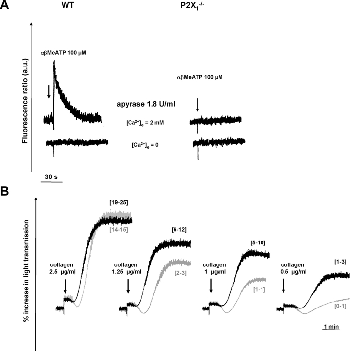
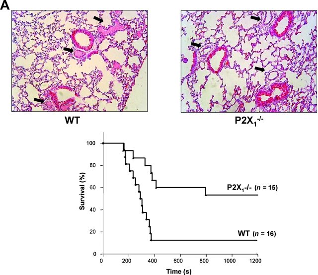
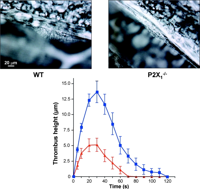
The P2X1 receptor is a fast ATP-gated cation channel expressed in blood platelets, where its role has been difficult to assess due to its rapid desensitization and the lack of pharmacological tools. In this paper, we have used P2X1-/- and wild-type mouse platelets, treated with apyrase to prevent desensitization, to demonstrate the function of P2X1 in the response to thrombogenic stimuli. In vitro, the collagen-induced aggregation and secretion of P2X1-deficient platelets was decreased, as was adhesion and thrombus growth on a collagen-coated surface, particularly when the wall shear rate was elevated. In vivo, the functional role of P2X1 could be demonstrated using two models of platelet-dependent thrombotic occlusion of small arteries, in which blood flow is characterized by a high shear rate. The mortality of P2X1-/- mice in a model of systemic thromboembolism was reduced and the size of mural thrombi formed after a laser-induced vessel wall injury was decreased as compared with normal mice, whereas the time for complete thrombus removal was shortened. Overall, the P2X1 receptor appears to contribute to the formation of platelet thrombi, particularly in arteries in which shear forces are high.


Figures

Figure 1.
Figure 1.
Tail bleeding time of WT and P2X1 −/− male mice. The bleeding time was normal in most of the P2X1 −/− mice (n = 34, median 107 s) as compared with WT mice (n = 32, median 85 s).
Figure 2.
Figure 2.
In vitro evaluation of platelet P2X1 receptor function. (A) [Ca2+]i measurements in washed platelet suspensions. In the presence of 1.8 U/ml apyrase, 100 μM αβMeATP elicited a [Ca2+]i rise in WT but not in P2X1 −/− platelets, which was abolished in the absence of external calcium (0.2 mM EGTA). (B) Aggregation of WT (black lines) and P2X1 −/− (gray lines) platelets in response to various concentrations of collagen. P2X1 −/− platelets showed a diminished response to collagen as compared with WT platelets. The percentage [3H]serotonin secretion, as indicated in brackets, was reduced in P2X1 −/− platelets as compared with WT platelets (values are from two independent experiments). Tracings are from one experiment representative of five independent experiments with identical results.
Figure 3.
Figure 3.
Role of the platelet P2X1 receptor in thrombus growth on a collagen-coated surface at high shear rates. (A) Surface coverage by platelet thrombi formed on a surface coated with 0.1 mg/ml type I collagen fibrils. Blood was perfused for 90–240 s at the indicated shear rates. Surface coverage by thrombi was significantly decreased in P2X1 −/− blood only at the shear rate of 6,000 s−1 (*, P = 0.032, n = 15). (B) Volume of thrombi formed on a 0.1-mg/ml collagen-coated surface after perfusion for 150 s at 6,000 s−1 wall shear rate with either WT or P2X1 −/− blood (P = 0.36, n = 6). Two single frame images at low magnification from a real time recording of one such experiment are also shown. Thrombi appeared as bright objects rendered fluorescent by the incorporation of mepacrine into platelet granules.
Figure 4.
Figure 4.
Resistance of P2X1 −/− mice to acute systemic intravascular thromboembolism. (A) Representative histological sections of lungs of WT or P2X1 −/− mice challenged with collagen and adrenaline (top). Frequent occurrence of occlusive intravascular thrombi in lungs of WT mice (left, arrows) compared with unobstructed vessels in lungs of P2X1 −/− mice (right, arrows). Time from 0.3 mg/kg collagen and 60 μg/kg adrenaline injection to death of mice (bottom). Results are expressed as the percentage of mice alive as a function of time. The effect of genotype on survival was significant by log-rank test (P = 0.0026). (B) Mechanical thromboembolism induced by i.v. infusion of hardened rat RBCs. 100 mg/kg nifedipine was administered per os 3 h before i.v. challenge with hardened RBCs. The effect of nifedipine treatment on survival of WT mice was significant by log-rank test ( P = 0.0003) as was the survival of nifedipine-treated WT mice as compared with P2X1 −/− mice (P = 0.0076). In contrast, the survival of WT mice was not significantly different as compared with that of P2X1 −/− mice (NS, P = 0.3909).
Figure 4.
Figure 4.
Resistance of P2X1 −/− mice to acute systemic intravascular thromboembolism. (A) Representative histological sections of lungs of WT or P2X1 −/− mice challenged with collagen and adrenaline (top). Frequent occurrence of occlusive intravascular thrombi in lungs of WT mice (left, arrows) compared with unobstructed vessels in lungs of P2X1 −/− mice (right, arrows). Time from 0.3 mg/kg collagen and 60 μg/kg adrenaline injection to death of mice (bottom). Results are expressed as the percentage of mice alive as a function of time. The effect of genotype on survival was significant by log-rank test (P = 0.0026). (B) Mechanical thromboembolism induced by i.v. infusion of hardened rat RBCs. 100 mg/kg nifedipine was administered per os 3 h before i.v. challenge with hardened RBCs. The effect of nifedipine treatment on survival of WT mice was significant by log-rank test ( P = 0.0003) as was the survival of nifedipine-treated WT mice as compared with P2X1 −/− mice (P = 0.0076). In contrast, the survival of WT mice was not significantly different as compared with that of P2X1 −/− mice (NS, P = 0.3909).
Figure 5.
Figure 5.
A key role of the platelet P2X1 receptor in arterial thrombosis. Representative photographs of the thrombus formed after laser-induced injury of the wall of mesenteric arterioles in WT and P2X1 −/− mice (top). Arterioles with a diameter of 65–100 μm were targets for injury. Kinetics of thrombus formation in mesenteric arterioles of WT (closed squares) and P2X1 −/− (closed triangles) mice (top). Values are the mean thrombus height (±SEM) at each time point in four WT mice (n = 15 vessels) and four P2X1 −/− mice (n = 16 vessels), and time 0 corresponds to the time of injury.

References

    1. Gachet, C. 2001. ADP receptors of platelets and their inhibition. Thromb. Haemost. 86:222–232. - PubMed
    1. Léon, C., B. Hechler, M. Freund, A. Eckly, C. Vial, P. Ohlmann, A. Dierich, M. LeMeur, J.P. Cazenave, and C. Gachet. 1999. Defective platelet aggregation and increased resistance to thrombosis in purinergic P2Y(1) receptor-null mice. J. Clin. Invest. 104:1731–1737. - PMC - PubMed
    1. Fabre, J.E., M. Nguyen, A. Latour, J.A. Keifer, L.P. Audoly, T.M. Coffman, and B.H. Koller. 1999. Decreased platelet aggregation, increased bleeding time and resistance to thromboembolism in P2Y1-deficient mice. Nat. Med. 5:1199–1202. - PubMed
    1. Hollopeter, G., H. Jantzen, D. Vincent, G. Li, L. England, V. Ramakrishan, R. Yang, P. Nurden, A. Nurden, D. Julius, and P. Conley. 2001. Identification of the platelet ADP receptor targeted by antithrombotic drugs. Nature. 409:202–207. - PubMed
    1. Foster, C.J., D.M. Prosser, J.M. Agans, Y. Zhai, M.D. Smith, J.E. Lachowicz, F.L. Zhang, E. Gustafson, F.J. Monsma, Jr., M.T. Wiekowski, et al. 2001. Molecular identification and characterization of the platelet ADP receptor targeted by thienopyridine antithrombotic drugs. J. Clin. Invest. 107:1591–1598. - PMC - PubMed

Publication types

MeSH terms