. 2003 Aug 18;198(4):557-67.
doi: 10.1084/jem.20030788. Epub 2003 Aug 11.

Identification of PVR (CD155) and Nectin-2 (CD112) as cell surface ligands for the human DNAM-1 (CD226) activating molecule

Affiliations

Identification of PVR (CD155) and Nectin-2 (CD112) as cell surface ligands for the human DNAM-1 (CD226) activating molecule

Cristina Bottino et al. J Exp Med. .

Abstract

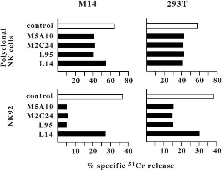
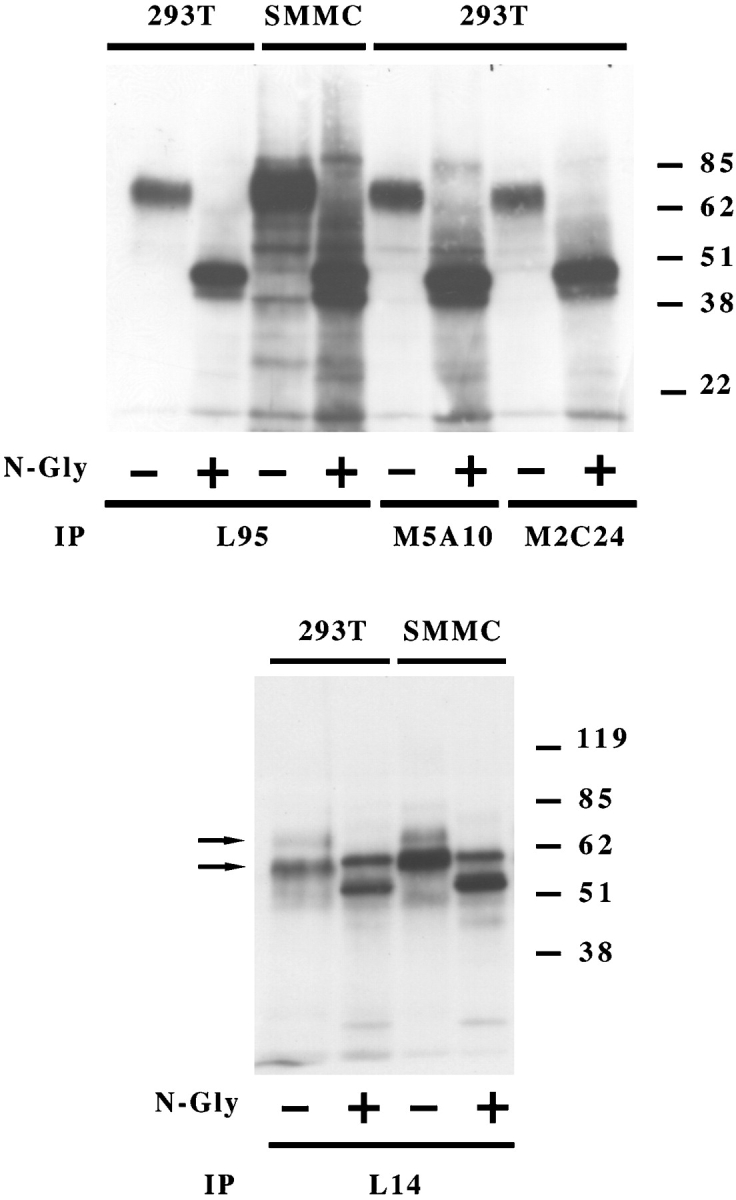
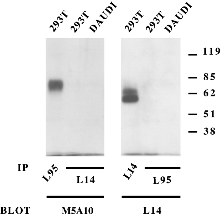
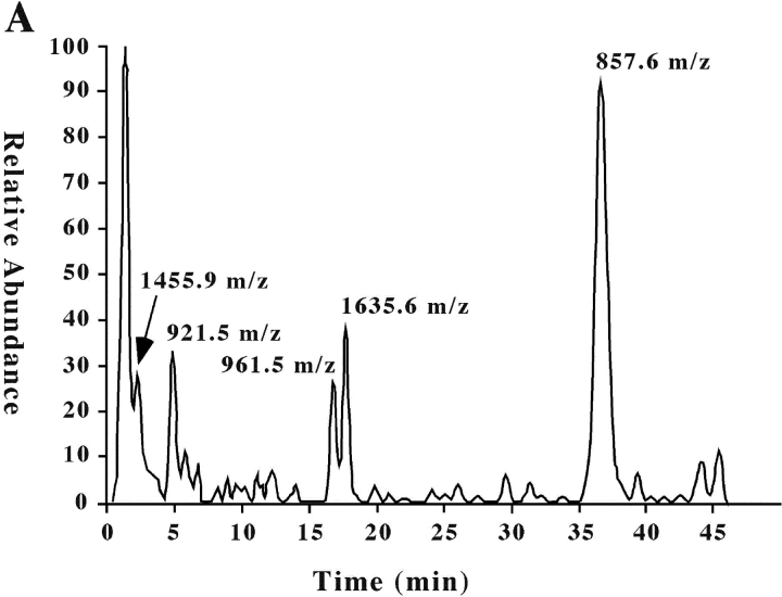
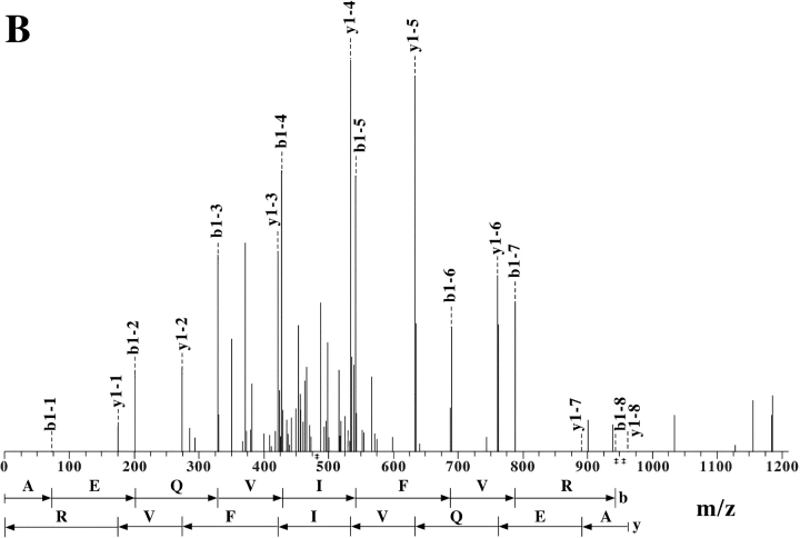
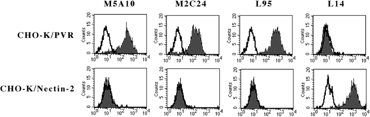
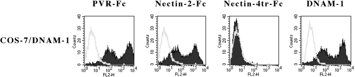
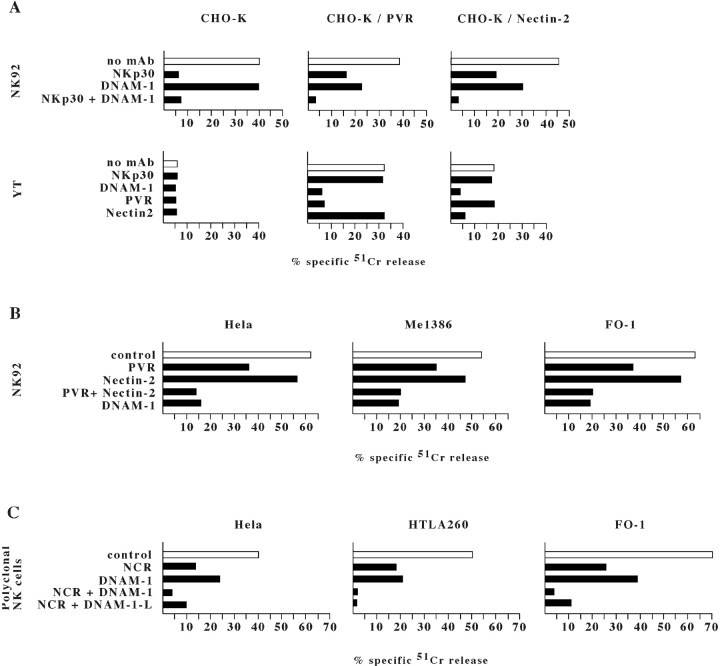
Human natural killer (NK) cells express a series of activating receptors and coreceptors that are involved in recognition and killing of target cells. In this study, in an attempt to identify the cellular ligands for such triggering surface molecules, mice were immunized with NK-susceptible target cells. On the basis of a functional screening, four mAbs were selected that induced a partial down-regulation of the NK-mediated cytotoxicity against the immunizing target cells. As revealed by biochemical analysis, three of such mAbs recognized molecules of approximately 70 kD. The other mAb reacted with two distinct molecules of approximately 65 and 60 kD, respectively. Protein purification followed by tryptic digestion and mass spectra analysis, allowed the identification of the 70 kD and the 65/60 kD molecules as PVR (CD155) and Nectin-2 delta/alpha (CD112), respectively. PVR-Fc and Nectin-2-Fc soluble hybrid molecules brightly stained COS-7 cells transfected with the DNAM-1 (CD226) construct, thus providing direct evidence that both PVR and Nectin-2 represent specific ligands for the DNAM-1 triggering receptor. Finally, the surface expression of PVR or Nectin-2 in cell transfectants resulted in DNAM-1-dependent enhancement of NK-mediated lysis of these target cells. This lysis was inhibited or even virtually abrogated upon mAb-mediated masking of DNAM-1 (on NK cells) or PVR or Nectin-2 ligands (on cell transfectants).


Figures

Figure 1.
Figure 1.
Inhibition of NK-mediated cytotoxicity by M5A10, M2C24, L95, and L14 mAbs. A polyclonal NK cell population (top panels) or the NK92 NK cell line (bottom panels) pretreated with anti-NKG2D mAb (BAT221; for 15 min at room temperature) were analyzed for cytolytic activity against the indicated target cell lines in the presence of either a control mAb (white bars) or one or another of the indicated mAbs (black bars). The effector/target ratio used was 10:1 for polyclonal NK cells and 20:1 for NK92. The results are representative of three independent experiments; the standard deviation of the mean of the triplicates was <5%.
Figure 2.
Figure 2.
Biochemical characterization of the surface molecules recognized by M5A10, M2C24, L95, and L14 mAbs. The indicated cell lines were surface labeled with biotin and immunoprecipitated with L95, M5A10, M2C24 (top panel), and L14 (bottom panel) mAbs. Samples were treated (+) or not (−) with N-glycosidase F and analyzed in an 8% SDS-PAGE under reducing conditions. Molecular weight markers (kD) are indicated.
Figure 3.
Figure 3.
Western blot analysis of M5A10, L95, and L14 mAbs. The indicated cell lines were immunoprecipitated with L95 and L14 mAbs. Samples were analyzed in an 8% SDS-PAGE under nonreducing conditions and probed with biotin-labeled M5A10 and L14 mAbs. Molecular weight markers (kD) are indicated. DAUDI cell lines were used as negative control.
Figure 4.
Figure 4.
Extract ion chromatogram of the L14-antigen tryptic digest and representative fragmentation spectrum for ion m/z 961. (A) Ion chromatogram displaying the base peaks of the tryptic digest of L14-specific molecule analyzed on a LC-MS/MS system as described in Materials and Methods. (B) MS/MS of the peptide with molecular ion 961.55 m/z from which the amino acid sequence AEQVIFVR was obtained in the automated system from NH2 to COOH terminal and vice versa.
Figure 4.
Figure 4.
Extract ion chromatogram of the L14-antigen tryptic digest and representative fragmentation spectrum for ion m/z 961. (A) Ion chromatogram displaying the base peaks of the tryptic digest of L14-specific molecule analyzed on a LC-MS/MS system as described in Materials and Methods. (B) MS/MS of the peptide with molecular ion 961.55 m/z from which the amino acid sequence AEQVIFVR was obtained in the automated system from NH2 to COOH terminal and vice versa.
Figure 5.
Figure 5.
Surface reactivity of M5A10, M2C24, L95 and L14 mAbs with CHO-K cells transfected with the PVR or Nectin-2 constructs CHO-K cells transfected with the PVRα or Nectin-2δ (or Nectin-2α, data not shown) constructs were stained with the indicated mAbs followed by PE-conjugated goat anti–mouse isotype-specific second reagent and analyzed by flow cytometry. White profiles indicate cells incubated with the second reagent only.
Figure 6.
Figure 6.
Surface reactivity of PVR-Fc or Nectin-2-Fc molecules with DNAM-1 cell transfectants COS-7 cells transfected with DNAM-1 construct were stained with PVR-Fc, Nectin-2-Fc, or Nectin-4tr-Fc (negative control) molecules, followed by PE-conjugated goat anti–human second reagent, or with GN18 (anti-DNAM-1) mAb, followed by PE-conjugated goat anti–mouse isotype-specific second reagent. Samples were analyzed by flow cytometry. White profiles indicate cells incubated with the second reagent only.
Figure 7.
Figure 7.
mAb-mediated disruption of DNAM-1/ligands interaction inhibits the NK-mediated lysis of PVR or Nectin-2 cell transfectants and of tumor cell targets. (A) NK92 or YT NK cell lines were analyzed for cytolytic activity against the CHO-K cells either untransfected or transfected with PVRα or Nectin-2δ constructs. Cytolytic assays were performed either in the absence of mAb (white bars) or in the presence of F252, GN18, L14, and L95 mAbs to the indicated molecules (black bars). The effector/target ratio used was10:1 for NK92 and 40:1 for YT. Similar results were obtained with Nectin-2α transfectants (data not shown). (B) NK92 NK cell line pretreated with anti-NKG2D mAb (for 15 min at room temperature) was analyzed for cytolytic activity against the indicated tumor target cells in the presence of either a control mAb (white bars) or mAbs (as in panel A) to the indicated molecules (black bars). The effector/target ratio used was 10:1. (C) A polyclonal NK cell population pretreated with anti-NKG2D mAb was analyzed for cytolytic activity against the indicated tumor target cells in the presence of either a control mAb (white bars) or mAbs to NCR (F252 and KL247), DNAM-1 (KRA236), or DNAM-1-Ligands (L95 and L14). These mAbs were used alone or in combination as indicated. The effector/target ratio used was 4:1. The results are representative of four independent experiments; the standard deviation of the mean of the triplicates was <4%.

References

    1. Moretta, A., C. Bottino, M. Vitale, D. Pende, R. Biassoni, M.C. Mingari, and L. Moretta. 1996. Receptors for HLA-class I molecules in human natural killer cells. Annu. Rev. Immunol. 14:619–648. - PubMed
    1. Long, E.O. 1999. Regulation of immune responses through inhibitory receptors. Annu. Rev. Immunol. 17:875–904. - PubMed
    1. Ravetch, J.V., and L.L. Lanier. 2000. Immune inhibitory receptors. Science. 290:84–89. - PubMed
    1. Shilling, H.G., N. Young, L.A. Guethlein, C.N. Wheng, C.M. Gardiner, D. Tyan, and P. Parham. 2002. Genetic control of human NK cell repertoire. J. Immunol. 169:239–247. - PubMed
    1. Moretta, A., C. Bottino, M. Vitale, D. Pende, C. Cantoni, M.C. Mingari, R. Biassoni, and L. Moretta. 2001. Activating receptors and co-receptors involved in human natural killer cell-mediated cytolysis. Annu. Rev. Immunol. 19:197–223. - PubMed

Publication types

MeSH terms