. 2003 Aug 18;162(4):731-41.
doi: 10.1083/jcb.200304031. Epub 2003 Aug 11.

Spatial restriction of alpha4 integrin phosphorylation regulates lamellipodial stability and alpha4beta1-dependent cell migration

Affiliations

Spatial restriction of alpha4 integrin phosphorylation regulates lamellipodial stability and alpha4beta1-dependent cell migration

Lawrence E Goldfinger et al. J Cell Biol. .

Abstract

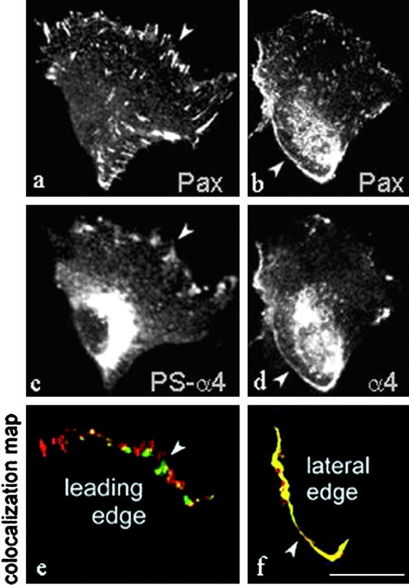
Integrins coordinate spatial signaling events essential for cell polarity and directed migration. Such signals from alpha4 integrins regulate cell migration in development and in leukocyte trafficking. Here, we report that efficient alpha4-mediated migration requires spatial control of alpha4 phosphorylation by protein kinase A, and hence localized inhibition of binding of the signaling adaptor, paxillin, to the integrin. In migrating cells, phosphorylated alpha4 accumulated along the leading edge. Blocking alpha4 phosphorylation by mutagenesis or by inhibition of protein kinase A drastically reduced alpha4-dependent migration and lamellipodial stability. alpha4 phosphorylation blocks paxillin binding in vitro; we now find that paxillin and phospho-alpha4 were in distinct clusters at the leading edge of migrating cells, whereas unphosphorylated alpha4 and paxillin colocalized along the lateral edges of those cells. Furthermore, enforced paxillin association with alpha4 inhibits migration and reduced lamellipodial stability. These results show that topographically specific integrin phosphorylation can control cell migration and polarization by spatial segregation of adaptor protein binding.


Figures

Figure 1.
Figure 1.
Characterization of a phospho-specific α-PSα4 mAb. (A) The α4 integrin–cytoplasmic tail recombinant proteins were incubated with protein kinase A (PKA) in the presence of [γ32P]ATP. The autoradiograph in the top panel shows that the α4 tail is efficiently phosphorylated by PKA. Only PKA-phosphorylated α4 tail protein shows reactivity with the α-PSα4 mAb. Coomassie stained gels are shown to indicate equal loading of α4 tail proteins. (B) Western blot of cell extracts prepared from Jurkat cells and JB4 cells, an α4-deficient Jurkat variant cell line, indicating specific reactivity of the α-PSα4 antibody with endogenous α4 integrin. (C) α4 integrins were immunoprecipitated from stably transfected CHO cells that had been surface labeled with biotin before lysis for detection of α4 protein using avidin-HRP. α-PSα4 antibody reacts with immunoprecipitated wt α4, but does not recognize α4 containing a Ser988 to Ala mutation.
Figure 2.
Figure 2.
α4 integrin is preferentially phosphorylated at the leading edge of migrating cells. (a–c) CHO cells expressing human α4 integrin were plated onto dishes coated with 2 μg/ml CS-1 (α4β1-binding) fragment of fibronectin and scratch wounded. The localization of the phosphorylated α4 was assessed by staining with α-PSα4. Serine phosphorylated α4 is localized to clusters at the leading edges of polarized cells, and in the perinuclear region. (d) Primary human peripheral blood monocytes migrating on CS-1 show leading edge localization of phosphorylated endogenous α4. Bar, 25 μm. (e) A7r5 rat smooth muscle cells were plated on fibronectin on 3-μm porous filters by the method of Cho and Klemke (2002). Pseudopodia (Pd) and cell bodies (CB) were isolated, lysed and α4 was immunoprecipitated with HP2/1 mAb for α4. Immunoprecipitates were blotted with α-PSα4, then stripped and reprobed with a rabbit polyclonal antibody (Rb038) raised against α4. Phosphorylated α4 integrin is highly enriched in leading pseudopodia in polarized cells.
Figure 3.
Figure 3.
The phosphorylation-resistant α4(S988A) mutation inhibits α4-dependent cell migration. (A) CHO cells stably expressing wt α4 or α4(S988A) were plated onto dishes coated with 2 μg/ml CS-1, scratch wounded at confluence, and allowed to migrate into the wound space for 16 h. The ratio of the final wound area to the area immediately after scratching is indicated as percent closure. The cells bearing α4(S988A) manifest drastically reduced α4-dependent migration. (B) CHO cells expressing α4wt (a and b) or α4(S988A) cells (c and d) were fixed 16 h after scratch wounding and stained with antibodies to α4 (a and c) or with α-PSα4 (b and d). α4 is localized around the cell periphery, but is phosphorylated only at the leading edge. α-PSα4 does not react with CHO cells bearing α4(S988A). Bar, 25 μm.
Figure 4.
Figure 4.
α4 phosphorylation is required for cell polarization, lamellipodial stabilization, and directed migration. (A) CHO cells bearing α4 wt, α4(S988A), or α4 fused to paxillin, plated on dishes coated with 2 μg/ml CS-1, were observed in random migration assays by phase-contrast microscopy and photographed every 10 min for 4 h. Representative cells are shown at times 0, 60, 120, and 180 min. Persistence tracks indicate displacements of cell centroids over 240 min. CHO cells bearing α4 polarize, extend lamellipodia and migrate in the direction of the lamellipodium. Those bearing α4(S988A) or an α4-paxillin chimera do not polarize and do not migrate. (B) Migration rates are shown based on cell centroid assignments for at least 40 cells/trial, n = 3. Migration rates in the right panel indicate average cell displacement between successive time points. CHO cells bearing α4wt exhibit persistent, high migration rates over 4 h, whereas cells bearing α4(S988A) or an α4-paxillin chimera maintain significantly lower migration rates over the experimental time frame. (C) The number and persistence of protrusions was tracked by phase-contrast microscopy. CHOα4wt and CHOα4(S988A) cells plated on CS-1 were photographed every minute for 10 min, and protrusions were scored visually in successive images (n = 10 cells for each cell type). (B and C) Error bars represent SEM for all cells counted.
Figure 5.
Figure 5.
Inhibition of PKA activity blocks α4 phosphorylation at the leading edge and α4-dependent migration. (A) CHO cells bearing α4 were plated on dishes coated with 2 μg/ml CS-1 and confluent monolayers were scratch wounded, and incubated with 100 μM Rp-cAMP (b), 30 μM H-89 (c), or without inhibitors (a) for 30 min, and then fixed and stained with α-PSα4. Rp-cAMP or H-89 treatment eliminates phosphorylation of α4 at the leading edge. (B) CHO cells bearing α4 were plated on dishes coated with 2 μg/ml CS-1 (α4β1) or 3Fn(9-11) (α5β1) and confluent monolayers were scratch wounded, and incubated with or without Rp-cAMP at 37°C for 16 h. Migration into the wound was assessed as described in Materials and methods. Treatment with Rp-cAMP inhibits migration on CS-1, but has no effect on migration on the α5 integrin–binding 3Fn(9–11) fragment of fibronectin. Error bars are the SD from the average width of the wound space measured in three independent trials. (C) CHOα4wt and CHOα4(S988A) cells were plated on CS-1, allowed to reach confluence, and either left unscratched, or scratch wounded with multiple scratches in a grid pattern. Wounded and unwounded cultures were incubated for 30 min in the presence or absence of 30 μM H89, and then extracted in lysis buffer. Lysates were adjusted to identical protein concentrations and analyzed by SDS-PAGE followed by Western blotting with α-PSα4.
Figure 5.
Figure 5.
Inhibition of PKA activity blocks α4 phosphorylation at the leading edge and α4-dependent migration. (A) CHO cells bearing α4 were plated on dishes coated with 2 μg/ml CS-1 and confluent monolayers were scratch wounded, and incubated with 100 μM Rp-cAMP (b), 30 μM H-89 (c), or without inhibitors (a) for 30 min, and then fixed and stained with α-PSα4. Rp-cAMP or H-89 treatment eliminates phosphorylation of α4 at the leading edge. (B) CHO cells bearing α4 were plated on dishes coated with 2 μg/ml CS-1 (α4β1) or 3Fn(9-11) (α5β1) and confluent monolayers were scratch wounded, and incubated with or without Rp-cAMP at 37°C for 16 h. Migration into the wound was assessed as described in Materials and methods. Treatment with Rp-cAMP inhibits migration on CS-1, but has no effect on migration on the α5 integrin–binding 3Fn(9–11) fragment of fibronectin. Error bars are the SD from the average width of the wound space measured in three independent trials. (C) CHOα4wt and CHOα4(S988A) cells were plated on CS-1, allowed to reach confluence, and either left unscratched, or scratch wounded with multiple scratches in a grid pattern. Wounded and unwounded cultures were incubated for 30 min in the presence or absence of 30 μM H89, and then extracted in lysis buffer. Lysates were adjusted to identical protein concentrations and analyzed by SDS-PAGE followed by Western blotting with α-PSα4.
Figure 6.
Figure 6.
Paxillin colocalizes with nonphosphorylated α4 at the lateral and trailing edges but not with phospho-α4 at the leading edge. CHO cells expressing α4wt were plated on CS-1, scratch wounded and stained with antibodies to paxillin (a) and total α4 (b), or paxillin (c) and phospho-α4 (d). Images shown are confocal micrographs of 0.1-μm basal sections. Cells that are separated from the monolayer are shown in a through d to demonstrate colocalization of paxillin (red) with total α4 (green) at the lateral and trailing edges. Colocalization maps are shown in e and f. Yellow pseudocolor indicates overlap of red and green fluorescence. Percent colocalization in the indicated regions is shown in g. (h) Percent colocalization between paxillin and phospho-α4 or total α4 staining across the whole cell area, in 200 cells analyzed per case (t test = 1.15 × 10⁻⁸ significance). (g and h) Error bars represent SEM of colocalization of red and green per pixel. Bar, 25 μm.
Figure 6.
Figure 6.
Paxillin colocalizes with nonphosphorylated α4 at the lateral and trailing edges but not with phospho-α4 at the leading edge. CHO cells expressing α4wt were plated on CS-1, scratch wounded and stained with antibodies to paxillin (a) and total α4 (b), or paxillin (c) and phospho-α4 (d). Images shown are confocal micrographs of 0.1-μm basal sections. Cells that are separated from the monolayer are shown in a through d to demonstrate colocalization of paxillin (red) with total α4 (green) at the lateral and trailing edges. Colocalization maps are shown in e and f. Yellow pseudocolor indicates overlap of red and green fluorescence. Percent colocalization in the indicated regions is shown in g. (h) Percent colocalization between paxillin and phospho-α4 or total α4 staining across the whole cell area, in 200 cells analyzed per case (t test = 1.15 × 10⁻⁸ significance). (g and h) Error bars represent SEM of colocalization of red and green per pixel. Bar, 25 μm.
Figure 7.
Figure 7.
Colocalization of unphosphorylated α4 with paxillin. CHO cells stably expressing nonphosphorylatable α4(S988A) (a and c) or pseudo-phosphorylated α4(S988D) (b and d) were plated on CS-1 and scratch wounded. The cells were fixed and stained with antibodies to paxillin (red) and α4 (green). In a and c, the cells remain rounded. Paxillin and α4 are coclustered around the cell perimeters, and paxillin is also found in α4-deficient clusters at the basal surface. In (b and d), CHO cells expressing α4(S988D) are spread, and paxillin is localized in clusters throughout the basal surface and at the perimeter. However, α4(S988D) staining shows widespread basal localization but virtually no colocalization with paxillin. Bar, 25 μm.
Figure 8.
Figure 8.
Enforced association of paxillin with α4 integrin inhibits migration. (A) CHO cells expressing α4 or an α4-paxillin chimera, were plated onto dishes coated with 2 μg/ml CS-1, scratch wounded at confluence, and allowed to migrate into the wound space for 16 h. The ratio of the final wound area to the area immediately after scratching is indicated as percent closure. Direct paxillin fusion to α4 integrin drastically reduces α4-dependent migration. Error bars are the SD from the average width of the wound space measured in three independent trials. (B) α4 integrins were immunoprecipitated from stably transfected CHO cells that had been surface labeled with membrane- impermeable biotin before lysis. Surface α4 was detected by staining immunoblots with avidin-HRP, and phospho-α4 was detected by staining with α-PSα4. The antibody reacts with immunoprecipitated α4 and the α4-paxillin chimera but not α4(S988A). (C) α4-paxillin chimera–transfected CHO cells were plated on CS-1, allowed to spread, and fixed and stained for α4 (a, green) and paxillin (b, red); the indicated region is shown as overlay in (c). Paxillin colocalizes with α4 at the cell periphery, but is also present in clusters across the basal surface that do not contain α4. Bar, 25 μm.
Figure 8.
Figure 8.
Enforced association of paxillin with α4 integrin inhibits migration. (A) CHO cells expressing α4 or an α4-paxillin chimera, were plated onto dishes coated with 2 μg/ml CS-1, scratch wounded at confluence, and allowed to migrate into the wound space for 16 h. The ratio of the final wound area to the area immediately after scratching is indicated as percent closure. Direct paxillin fusion to α4 integrin drastically reduces α4-dependent migration. Error bars are the SD from the average width of the wound space measured in three independent trials. (B) α4 integrins were immunoprecipitated from stably transfected CHO cells that had been surface labeled with membrane- impermeable biotin before lysis. Surface α4 was detected by staining immunoblots with avidin-HRP, and phospho-α4 was detected by staining with α-PSα4. The antibody reacts with immunoprecipitated α4 and the α4-paxillin chimera but not α4(S988A). (C) α4-paxillin chimera–transfected CHO cells were plated on CS-1, allowed to spread, and fixed and stained for α4 (a, green) and paxillin (b, red); the indicated region is shown as overlay in (c). Paxillin colocalizes with α4 at the cell periphery, but is also present in clusters across the basal surface that do not contain α4. Bar, 25 μm.

Similar articles

Cited by

References

    1. Alt, A., M. Ohba, L. Li, M. Gartsbein, A. Belanger, M.F. Denning, T. Kuroki, S.H. Yuspa, and T. Tennenbaum. 2001. Protein kinase Cdelta-mediated phosphorylation of alpha6beta4 is associated with reduced integrin localization to the hemidesmosome and decreased keratinocyte attachment. Cancer Res. 61:4591–4598. - PubMed
    1. Arroyo, A.G., J.T. Yang, H. Rayburn, and R.O. Hynes. 1996. Differential requirements for α4 integrins in hematopoiesis. Cell. 85:997–1008. - PubMed
    1. Baker, S.E., O. Skalli, R.D. Goldman, and J.C. Jones. 1997. Laminin-5 and modulation of keratin cytoskeleton arrangement in FG pancreatic carcinoma cells: involvement of IFAP300 and evidence that laminin-5/cell interactions correlate with a dephosphorylation of alpha 6A integrin. Cell Motil. Cytoskeleton. 37:271–286. - PubMed
    1. Calderwood, D.A., R. Zent, R. Grant, D.J. Rees, R.O. Hynes, and M.H. Ginsberg. 1999. The Talin head domain binds to integrin beta subunit cytoplasmic tails and regulates integrin activation. J. Biol. Chem. 274:28071–28074. - PubMed
    1. Chan, B.M.C., P.D. Kassner, J.A. Schiro, H.R. Byers, T.S. Kupper, and M.E. Hemler. 1992. Distinct cellular functions mediated by different VLA integrin α subunit cytoplasmic domains. Cell. 68:1051–1060. - PubMed

Publication types