Review
. 2003 Sep 30;100 Suppl 1(Suppl 1):11830-5.
doi: 10.1073/pnas.1734203100. Epub 2003 Aug 11.

Stem cells of the skin epithelium

Affiliations
Review

Stem cells of the skin epithelium

Laura Alonso et al. Proc Natl Acad Sci U S A. .

Abstract

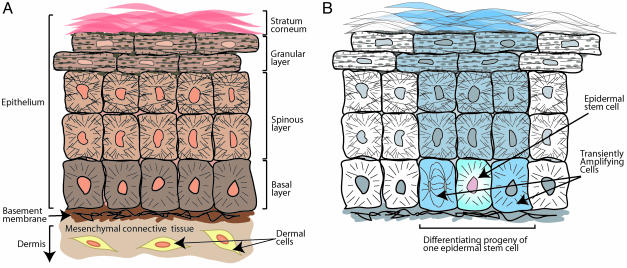
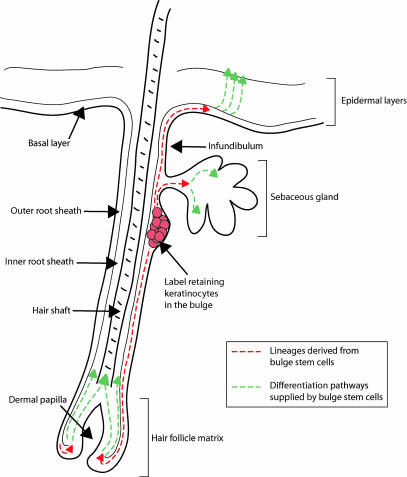
Tissue stem cells form the cellular base for organ homeostasis and repair. Stem cells have the unusual ability to renew themselves over the lifetime of the organ while producing daughter cells that differentiate into one or multiple lineages. Difficult to identify and characterize in any tissue, these cells are nonetheless hotly pursued because they hold the potential promise of therapeutic reprogramming to grow human tissue in vitro, for the treatment of human disease. The mammalian skin epithelium exhibits remarkable turnover, punctuated by periods of even more rapid production after injury due to burn or wounding. The stem cells responsible for supplying this tissue with cellular substrate are not yet easily distinguishable from neighboring cells. However, in recent years a significant body of work has begun to characterize the skin epithelial stem cells, both in tissue culture and in mouse and human skin. Some epithelial cells cultured from skin exhibit prodigious proliferative potential; in fact, for >20 years now, cultured human skin has been used as a source of new skin to engraft onto damaged areas of burn patients, representing one of the first therapeutic uses of stem cells. Cell fate choices, including both self-renewal and differentiation, are crucial biological features of stem cells that are still poorly understood. Skin epithelial stem cells represent a ripe target for research into the fundamental mechanisms underlying these important processes.


Figures

Fig. 1.
Fig. 1.
(A) Diagrammatic representation of skin epithelial histology. Cells of the basal layer attach to an underlying basement membrane. Basal cells are mitotically active, but they lose this potential when they detach from the basement membrane and embark on the outward trek toward the skin surface. As basal cells enter the spinous layer, they strengthen their cytoskeletal and intercellular connections, gaining resilience to mechanical stress. Once this task is completed, the cells enter the granular layer, where they produce the epidermal barrier. The barrier precursors consist of two major components: (i) glutamine- and lysine-rich cornified envelope precursor proteins, which are synthesized and deposited beneath the plasma membrane, and (ii) lamellar granules, which are filled with lipid bilayers. As the cells enter the final phases of terminal differentiation, a flux of calcium activates the enzyme transglutaminase, which biochemically cross-links the cornified envelope proteins through ε-(γ-glutamyl)lysine isopeptide bonds and which activates the extrusion of the lipid bilayers onto this scaffold. Cell death ensues, leaving dead, flattened squames at the skin surface, the end-process of terminal differentiation. These squames of the stratum corneum eventually slough from the skin surface, to be replenished continually by inner layer cells moving outward. (B) Diagram of the epidermal proliferative unit. A putative slow-cycling epidermal stem cell occasionally divides, giving rise to a stem cell daughter and a transiently amplifying daughter. The transiently amplifying cell divides two to four times, and these progeny then leave the basal layer and execute a program of terminal differentiation. This model is based on retroviral transduction of a β-galactosidase gene into cultured keratinocytes, which were then used in engraftments onto nude mice to trace stem cell lineages (9).
Fig. 2.
Fig. 2.
Diagram of the hair follicle and cell lineages supplied by epidermal stem cells. A compartment of multipotent stem cells is located in the bulge, which lies in the outer root sheath (ORS) just below the sebaceous gland. Contiguous with the basal layer of the epidermis, the ORS forms the external sheath of the hair follicle. The interior or the inner root sheath (IRS) forms the channel for the hair; as the hair shaft nears the skin surface, the IRS degenerates, liberating its attachments to the hair. The hair shaft and IRS are derived from the matrix, the transiently amplifying cells of the hair follicle. The matrix surrounds the dermal papilla, a cluster of specialized mesenchymal cells in the hair bulb. The multipotent stem cells found in the bulge are thought to contribute to the lineages of the hair follicle, sebaceous gland, and the epidermis (see red dashed lines). Transiently amplifying progeny of bulge stem cells in each of these regions differentiates as shown (see green dashed lines).

References

    1. Fuchs, E. & Cleveland, D. W. (1998) Science 279, 514-519. - PubMed
    1. Kalinin, A. E., Kajava, A. V. & Steinert, P. M. (2002) BioEssays 24, 789-800. - PubMed
    1. Gandarillas, A. (2000) Exp. Gerontol. 35, 53-62. - PubMed
    1. Potten, C. S. (1975) Br. J. Dermatol. 93, 649-658. - PubMed
    1. Rheinwald, J. G. & Green, H. (1975) Cell 6, 331-343. - PubMed

Publication types