. 2003 Aug 19;100(17):9756-61.
doi: 10.1073/pnas.1733910100. Epub 2003 Aug 11.

Protein grafting of an HIV-1-inhibiting epitope

Affiliations

Protein grafting of an HIV-1-inhibiting epitope

Samuel K Sia et al. Proc Natl Acad Sci U S A. .

Abstract

Protein grafting, the transfer of a binding epitope of one ligand onto the surface of another protein, is a potentially powerful technique for presenting peptides in preformed and active three-dimensional conformations. Its utility, however, has been limited by low biological activity of the designed ligands and low tolerance of the protein scaffolds to surface substitutions. Here, we graft the complete binding epitope (19 nonconsecutive amino acids with a solvent-accessible surface area of >2,000 A2) of an HIV-1 C-peptide, which is derived from the C-terminal region of HIV-1 gp41 and potently inhibits HIV-1 entry into cells, onto the surface of a GCN4 leucine zipper. The designed peptide, named C34coil, displays a potent antiviral activity approaching that of the native ligand. Moreover, whereas the linear C-peptide is unstructured and sensitive to degradation by proteases, C34coil is well structured, conformationally stable, and exhibits increased resistance to proteolytic degradation compared with the linear peptide. In addition to being a structured antiviral inhibitor, C34coil may also serve as the basis for the development of an alternative class of immunogens. This study demonstrates that "one-shot" protein grafting, without subsequent rounds of optimization, can be used to create ligands with structural conformations and improved biomedical properties.


Figures

Fig. 1.
Fig. 1.
Design of C34coil. (A) Superpositions of one C34 helix from the HIV-1 gp41 core (red) and one helix from the GCN4 leucine zipper (blue). The rest of gp41 is light gray, and the rest of GCN4 is dark gray. Shown are views down the 6-helix bundle of gp41 (Left) and down the superimposed helices (Right). The main-chain atoms of C34 (residues 632–657) and GCN4 (residues 4–29) superimpose to 1.2-Å rms deviation. (For the whole helix, from residues 629–659 of C34 and residues 1–31 of GCN4, the rms deviation is 1.7 Å due to helix fraying at the ends.) (B) Surface diagrams of the 6-helix bundle of HIV-1 gp41 with one C34 helix shown in ribbon format (backbone of C34 helix in red; Left) the dimeric GCN4 leucine zipper with one GCN4 helix shown in ribbon format (backbone of GCN4 helix in dark blue; Center), and a model of C34coil (Right). Both the C34 binding epitope (orange) and the GCN4 hydrophobic core (light blue) are incorporated into C34coil. The solvent-accessible surface area of the C34 binding epitope is 2,061 Å2 (see Materials and Methods for calculations). (C) Helical wheel representations of C34 (red), GCN4 (blue), and C34coil (red and blue). The four residues at the g′ position of C34 and the four residues at the e position of GCN4 are identical and are shown in italics. C34coil is a covalent heterodimer of two peptides, (Cys)C34-GCN4 and (Cys)GCN4, connected by means of a disulfide bond [(Cys) denotes the addition of a Cys-Gly-Gly sequence to the N terminus of the peptide]. The boxed residues of C34 (at a′, d′, e′, and g′ positions) and GCN4 (at a, d, e, and g positions) are incorporated into the C34-GCN4 peptide and are equivalent to the binding epitope highlighted in orange and blue, respectively, in B. (D) Sequences of the peptides C34 (red), GCN4 (blue), (Cys)C34-GCN4 (red and blue), and (Cys)GCN4 (blue). The four residues in common in all peptides are shown in italics. The C34-GCN4 peptide, which lacks the N-terminal Cys-Gly-Gly, was also synthesized. Ac, an acetylated N terminus; NH2, an amidated C terminus. Note that the GCN4 sequence used in this study corresponds to GCN4-p1 in previous studies (32). See Materials and Methods for more details on the peptide design.
Fig. 2.
Fig. 2.
Biophysical characterization of C34coil. (A) CD spectrum of C34, C34-GCN4, and C34coil. The guanidine hydrochloride used to solubilize C34-GCN4 precludes measurements at wavelengths <210 nm. Experimental conditions were: PBS at pH 7.0 at 25°C. C34coil exhibits a helical structure ([θ]222 value of –29,400 deg cm2·dmol1), whereas C34 (–3,800 deg cm2·dmol1) and the C34-GCN4 peptide (–12,400 deg cm2·dmol1) exhibit relatively unstructured conformations. (B) Guanidine hydrochloride denaturation of C34coil, as monitored by CD spectroscopy at 222 nm. The fitted curve to a two-state unfolding transition is shown as a black line. C34coil unfolds at a midpoint of 3.6 M guanidine hydrochloride, with a free energy of unfolding of 9.3 kcal/mol. (C) Apparent molecular weight (MW) of C34coil, as determined by gel filtration chromatography. Shown are the elution times of molecular weight standards (▵) and C34coil (•). Also shown is the best-fit line of apparent MW versus elution time from the molecular weight standards (black line). C34coil exhibits an apparent molecular mass of 7,000 ± 2,000 Da (expected molecular mass of 8,740 Da for a monomer), with no detectable aggregation. (D) Sensitivity to proteolytic degradation of C34coil and C34. Peptides are incubated with proteinase K at 37°C, and the reaction products are monitored by reverse-phase HPLC. The relative ratios of proteinase K account for differences in both the protease concentrations and incubation times. Shown are the HPLC chromatograms. C34coil is ≈1,000-fold more resistant to degradation by proteinase K than by C34.
Fig. 3.
Fig. 3.
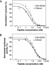
Inhibition of HIV-1 envelope-mediated membrane fusion. The data represent the mean ± SE of at least two separate experiments. (A) Inhibition of cell–cell fusion. The peptides were tested for inhibiting the fusion of HIV-1 Env-expressing cells (CHO gp160) with CD4-expressing cells (CD4 HeLa). The IC50 values for C34coil and C34-GCN4 are 3.1 ± 0.8 and 4.6 ± 0.9 nM, respectively. The (Cys)GCN4-homodimer shows no inhibitory activity up to 50 μM. The IC50 value of C34 in the cell–cell fusion assay, as reported in ref. , was 0.6 nM. (B) Inhibition of viral infectivity. The peptides were tested for inhibition of CD4-positive target cells (HOST4) by recombinant, luciferase-expressing HIV-1. The IC50 values for C34 coil and C34-GCN4 are 16 ± 2 and 19 ± 3 nM, respectively. The (Cys)GCN4-homodimer shows no inhibitory activity up to 50 μM. The IC50 value of C34 in the viral infectivity assay, as reported in ref. , was 2 nM.
Fig. 4.
Fig. 4.
Characterization of C34coil-N16K. (A) Unfolding of C34coil-N16K by guanidine hydrochloride as measured by CD spectroscopy. The midpoint of unfolding occurs at 3.0 M guanidine, with a free energy of unfolding of 9.0 kcal/mol. (B) Inhibition of HIV-1 entry by C34coil-N16K, as measured by the cell–cell fusion and viral infectivity assays. Standard deviations are shown as error bars. The IC50 values are 1.5 nM in the cell–cell fusion assay and 10 nM in the viral infectivity assay.

Similar articles

Cited by

References

    1. D'Souza, M. P., Cairns, J. S. & Plaeger, S. F. (2000) J. Am. Med. Assoc. 284, 215–222. - PubMed
    1. Eckert, D. M. & Kim, P. S. (2001) Annu. Rev. Biochem. 70, 777–810. - PubMed
    1. Moore, J. P. & Stevenson, M. (2000) Nat. Rev. Mol. Cell Biol. 1, 40–49. - PubMed
    1. Nabel, G. J. (2001) Nature 410, 1002–1007. - PubMed
    1. Wild, C. T., Shugars, D. C., Greenwell, T. K., McDanal, C. B. & Matthews, T. J. (1994) Proc. Natl. Acad. Sci. USA 91, 9770–9774. - PMC - PubMed

Publication types

MeSH terms

LinkOut - more resources