. 2003 Jul 23;3(1):12.
doi: 10.1186/1475-2867-3-12.

Inducible expression of catalytically active type 1 serine/threonine protein phosphatase in a human carcinoma cell line

Affiliations

Inducible expression of catalytically active type 1 serine/threonine protein phosphatase in a human carcinoma cell line

Jay E Reeder et al. Cancer Cell Int. .

Abstract

BACKGROUND: One of the major cellular serine/threonine protein phosphatases is protein phosphatase type 1 (PP1). Studies employing many eukaryotic systems all point to a crucial role for PP1 activity in controlling cell cycle progression. One physiological substrate for PP1 appears to be the product of the retinoblastoma susceptibility gene (pRB), a demonstrated tumor suppressor. The growth suppressive activity of pRB is regulated by its phosphorylation state. Of critical importance is the question of the in vivo effect of PP1 activity on pRB and growth regulation. As a first step towards addressing this question, we developed an inducible PP1 expression system to investigate the regulation of PP1 activity. RESULTS: We have established a cell line for inducing protein expression of the type 1, alpha-isotype, serine/threonine protein phosphatase (PP1alpha). A plasmid encoding a fusion protein of the catalytic subunit of PP1alpha with a 6-histidine peptide (6His) and a peptide from hemagluttinin (HA) was transfected into the UMUC3 transitional cell carcinoma cell line, previously transfected with the reverse tetracycline transactivator plasmid pUHD172-1neo. A stable cell line designated LLWO2F was established by selection with hygromycin B. 6His-HA-PP1alpha protein appeared in cell lysates within two hours following addition of doxycycline to the culture medium. This protein localizes to the nucleus as does endogenous PP1alpha, and was shown to associate with PNUTS, a PP1-nuclear targeting subunit. Like endogenous PP1alpha, immunocomplexed 6His-HA-PP1alpha is active toward phosphorylase a and the product of the retinoblastoma susceptibility gene, pRB. When forcibly overexpressing 6His-HA-PP1alpha, there is a concomitant decrease in endogenous PP1alpha levels. CONCLUSIONS: These data suggest the existence of an autoregulatory mechanism by which PP1alpha protein levels and activity remain relatively constant. RT-PCR analyses of isolated polysome fractions support the notion that this putative autoregulatory mechanism is exerted, at least in part, at the translational level. Implications of these findings for the study of PP1alpha function in vivo are discussed.


Figures

Figure 1
Figure 1
The 6His-HA-PP1α-pTEP4m plasmid. The plasmid contains the 6His-HA-PP1α fusion gene under the control of the TetO/CMV promoter. This promoter is active in the presence of the rtTA-nls protein and doxycycline. The plasmid contains an ampicillin resistance gene (Ampr) for selection in bacteria and the hygromycin resistance gene for selection in eukaryotic cells.
Figure 2
Figure 2
A. Doxycycline has no effect on PP1α protein expression in control cell lines. Equal quantities (50 ug) of whole-cell lysates harvested in the absence (indicated above the lanes with a (-) symbol), or presence (at 24 hr following addition, indicated above the lanes with a (+) symbol), of doxycycline were separated by SDS-PAGE and subjected to immunoblotting using antibody specific for PP1α. UMUC3 – parent cell line from which stable expressor LLWO2F was selected; LLWO1 – cell line generated from UMUC3 which was stably transfected with the reverse tetracycline transactivator plasmid pUHD172-1neo only. B – Doxycycline induces a protein in LLW02F cells that is recognized by antibody to hemagglutinin. Equal quantities (50 ug) of whole-cell lysates harvested at the various time points in hours after doxycycline addition (indicated above each lane) were separated by SDS-PAGE and subjected to immunoblotting as described in Materials and Methods. C – Time-course of recovery from doxycycline induction. LLWO2F cells were induced for 24 hr. At the end of this time, equal quantities (50 ug) of whole-cell lysates harvested at the various time points in hours following removal of doxycycline (indicated above each lane) were separated by SDS-PAGE and subjected to immunoblotting with antibody to hemagglutinin.
Figure 3
Figure 3
Immunohistochemistry with the antibody to hemagglutinin shows doxycycline-dependent expression of 6His-HA-PP1α in vivo. LLWO2F cells were grown in 12 well tissue culture plates and induced for 24 hours prior to fixation and staining as described in Materials and Methods. Panel A – Induced; Panel B – Uninduced.
Figure 4
Figure 4
Coprecipitation of 6His-HA-PP1α with GST-PNUTS. Doxycycline-induced LLWO2F cell lysates were mixed with either GST or GST-PNUTS bound to glutathione-Sepharose beads, washed, and the bound proteins separated by SDS-PAGE and then immunoblotted with antibody to hemagglutinin. Position of 6His-HA-PP1α is indicated to the left of the panel, and is present only in the GST-PNUTS lane. The band above 6His-HA-PP1α, which is also present in the GST-alone lane, results from non-specific reactivity with the secondary antibody (horse-radish peroxidase-conjugated anti-IgG) used for chemiluminescent detection
Figure 5
Figure 5
Dephosphorylation of phosphorylase a by anti-hemagglutinin immunoprecipitates from uninduced (Control) and induced (Doxycycline) LLW02 cells. Activity was assayed by the release of [32P] H3PO4 from radiolabeled phosphorylase a (1–2 × 10⁵ cpm/nmol). 1 unit of activity releases 1 nmol of phosphate/min at 30°C.
Figure 6
Figure 6
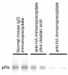
Dephosphorylation of pRB by anti-hemagglutinin immunoprecipitates from induced LLW02 cells. Samples were run in duplicate. Normal mouse IgG immunoprecipitate was used as a negative control. Anti-hemagglutinin immunoprecipitate, reaction run in the presence of okadaic acid (100 nM), a demonstrated PP1 inhibitor, was an additional negative control. Anti-hemagglutinin immunoprecipitate, revealing a decrease in 32P-labeled pRB band intensity, indicating removal of phosphate groups by 6His-HA-PP1α.
Figure 7
Figure 7
PP1 activity in lysates of cells expressing 6His-HA-PP1α. Cells were cultured for 24 hr either in the absence (closed diamonds) or presence (closed squares) of doxycycline. PP1 was assayed using the indicated amounts of lysate/assay. 5 nM okadaic acid was used to inhibit PP2A during the assay. 1 unit of PP1 activity releases 1 nmol of Pi/min at 30°C.
Figure 8
Figure 8
Time course of induction followed by western blotting of whole cell lysates using antibody to PP1α. Equal quantities (50 ug) of whole-cell lysates harvested at the various time points in hours after doxycycline addition (indicated above each lane) were separated by SDS-PAGE and subjected to immunoblotting using antibody specific for PP1α. Positions of endogenous PP1α and 6His-HA-PP1α are indicated by arrows to the left and right of the figure, respectively.
Figure 9
Figure 9
PP1α isoform-specific antibody immunoprecipitation and detection of endogenous and induced PP1α by western blotting. PP1α isoform-specific antibody was used to immunoprecipitate proteins from equal quantities (50 ug) of uninduced (first lane from the left, top panel) and induced LLWO2F cell lysate (second lane from the left, top panel). The precipitated proteins were then separated by SDS-PAGE and immunoblotted with the same isoform-specific antibody. The positions of 6His-HA-PP1α and PP1α are indicated to the left of the top panel. Whole-cell lysates (10 ug) were separated in the next two lanes. Last lane contains 1 ug of purified recombinant PP1α as a control for endogenous PP1α (37 kDa). Bottom panel shows identical experiment using antibody to hemagglutinin for immunoprecipitation and immunodetection. Although low levels of 6His-HA-PP1α can be detected in cell lysate from uninduced cells following western blotting, this protein is almost undetectable in the same lysate following immunoprecipitation and western blotting. This observation is most likely due to low efficiency of immunoprecipitation.
Figure 10
Figure 10
Isoform-specific antibody detection of PP1 by western blotting induced and uninduced LLWO2F whole-cell lysates. Different amounts (5 ug for the first and third lanes, 10 ug for the second and fourth lanes) of lysate from induced and uninduced cells were separated by SDS-PAGE in parallel and then immunoblotted using PP1α-, PP1δ-, and PP1γ1-specific antibody. Results illustrate downregulation of endogenous PP1α in response to increasing amounts of induced 6His-HA-PP1α, while the levels of PP1δ and PP1γ1 remain relatively constant. Lane C – control lane of isoform-specific antibody immunoprecipitation of respective isoforms from 50 ug of uninduced cell lysate.
Figure 11
Figure 11
RT-PCR of polysome and total RNA specific for endogenous PP1α and 6His-HA-PP1α. PCR products from polysome and total RNA from induced and uninduced cell lysates were prepared and analyzed as described in Materials and Methods. MW – molecular mass standard ladder.

Similar articles

Cited by

References

    1. Cohen P. The structure and regulation of protein phosphatases. Ann Rev Biochem. 1989;58:453–508. doi: 10.1146/annurev.bi.58.070189.002321. - DOI - PubMed
    1. Ingebritsen TS, Cohen P. Protein phosphatases: properties and role in cellular regulation. Science. 1983;221:331–338. - PubMed
    1. Shima H, Hatano Y, Chun YS, Sugimura T, Zhang Z, Lee EY, Nagao M. Identification of PP1 catalytic subunit isotypes PP1 gamma 1, PP1 delta and PP1 alpha in various rat tissues. Biochem Biophys Res Commun. 1993;192:1289–1296. doi: 10.1006/bbrc.1993.1556. - DOI - PubMed
    1. Cyert MS, Thorner J. Putting it on and taking it off: phosphoprotein phosphatase involvement in cell cycle regulation. Cell. 1989;57:891–893. - PubMed
    1. Mumby MC, Walter G. Protein serine/threonine phosphatases: structure regulation, and functions in cell growth. Physiol Rev. 1993;73:673–699. - PubMed