Comparative Study
. 2003 Aug 12;4(1):34.
doi: 10.1186/1471-2164-4-34.

Application of comparative genomics in the identification and analysis of novel families of membrane-associated receptors in bacteria

Affiliations
Comparative Study

Application of comparative genomics in the identification and analysis of novel families of membrane-associated receptors in bacteria

Vivek Anantharaman et al. BMC Genomics. .

Abstract

Background: A great diversity of multi-pass membrane receptors, typically with 7 transmembrane (TM) helices, is observed in the eukaryote crown group. So far, they are relatively rare in the prokaryotes, and are restricted to the well-characterized sensory rhodopsins of various phototropic prokaryotes.

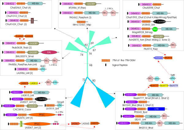
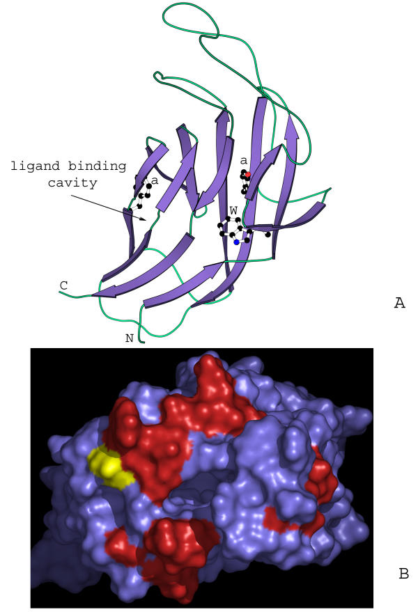
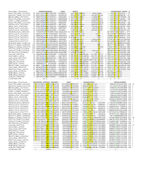
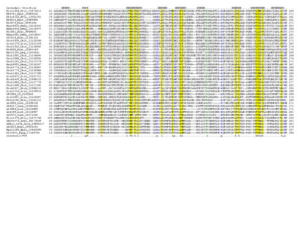
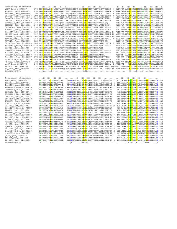
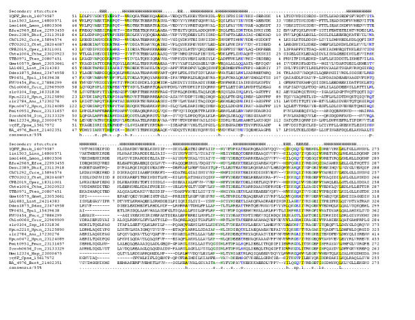
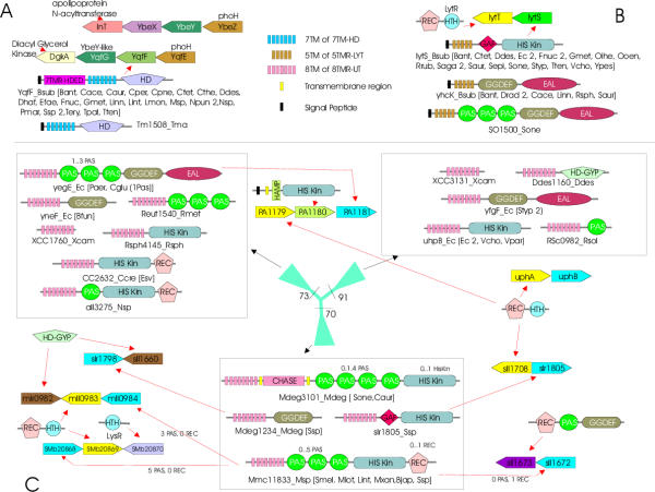
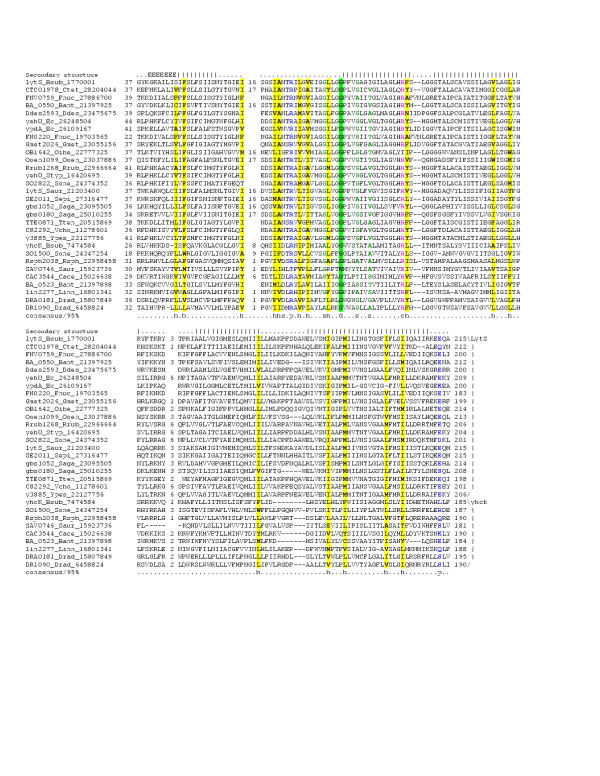
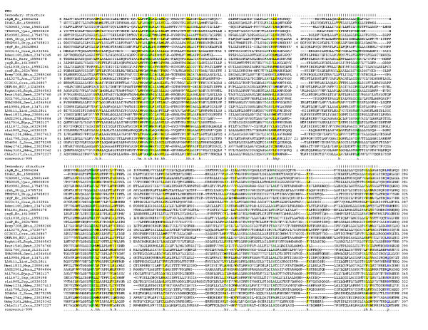
Results: Utilizing the currently available wealth of prokaryotic genomic sequences, we set up a computational screen to identify putative 7 (TM) and other multi-pass membrane receptors in prokaryotes. As a result of this procedure we were able to recover two widespread families of 7 TM receptors in bacteria that are distantly related to the eukaryotic 7 TM receptors and prokaryotic rhodopsins. Using sequence profile analysis, we were able to establish that the first members of these receptor families contain one of two distinct N-terminal extracellular globular domains, which are predicted to bind ligands such as carbohydrates. In their intracellular portions they contain fusions to a variety of signaling domains, which suggest that they are likely to transduce signals via cyclic AMP, cyclic diguanylate, histidine phosphorylation, dephosphorylation, and through direct interactions with DNA. The second family of bacterial 7 TM receptors possesses an alpha-helical extracellular domain, and is predicted to transduce a signal via an intracellular HD hydrolase domain. Based on comparative analysis of gene neighborhoods, this receptor is predicted to function as a regulator of the diacylglycerol-kinase-dependent glycerolipid pathway. Additionally, our procedure also recovered other types of putative prokaryotic multi-pass membrane associated receptor domains. Of these, we characterized two widespread, evolutionarily mobile multi-TM domains that are fused to a variety of C-terminal intracellular signaling domains. One of these typified by the Gram-positive LytS protein is predicted to be a potential sensor of murein derivatives, whereas the other one typified by the Escherichia coli UhpB protein is predicted to function as sensor of conformational changes occurring in associated membrane proteins

Conclusions: We present evidence for considerable variety in the types of uncharacterized surface receptors in bacteria, and reconstruct the evolutionary processes that model their diversity. The identification of novel receptor families in prokaryotes is likely to aid in the experimental analysis of signal transduction and environmental responses of several bacteria, including pathogens such as Leptospira, Treponema, Corynebacterium, Coxiella, Bacillus anthracis and Cytophaga.


Figures

Figure 1
Figure 1
Multiple sequence alignment of the 7TM domains of the 7TMR-DISM family. Multiple sequence alignment the 7TMR-DISM family was constructed using T-Coffee [73] after parsing high-scoring pairs from PSI-BLAST search results. The PHD-secondary structure [78] is shown above the alignment with | representing an α-helix. The 85% consensus shown below the alignment was derived using the following amino acid classes: hydrophobic (h: ALICVMYFW, yellow shading); small (s: ACDGNPSTV, green) and polar (p: CDEHKNQRST, blue). The limits of the domains are indicated by the residue positions, on each end of the sequence. The two major groups of these receptors, typically associated with either 7TMR-DISMED2s (upper group) or 7TMR-DISMED1s (lower group), are separated by a spacer. The numbers within the alignment are non-conserved inserts that have not been shown. The sequences are denoted by their gene name followed by the species abbreviation and GenBank Identifier (gi). The species abbreviations are as provided in Table 1. This alignment is provided as an additional file in the MS-WORD format (additional file 1).
Figure 2
Figure 2
Phylogenetic tree, domain architectures and gene neighborhoods of the 7TMR-DISM family. Phylogenetic relationships of the 7TMR-DISM domain containing proteins along with the domain architectures are shown. The seed alignment used for constructing the tree was one similar to that shown in Fig. 1. The RELL bootstrap values for the major branches are shown at their base. The thickness of a given branch is approximately proportional to the number of proteins contained within it. Domain architectures of the proteins in each branch of the tree are shown in boxes pointed to by the black arrows. The phyletic pattern of each family is shown, along with the number of proteins (if there are more than one). The gene neighborhood data for some of the genes encoding 7TMR-DISM encoding genes is depicted using block arrows. A red arrow indicates the domain architectures of proteins encoded by each gene. The species abbreviations are as shown in Table 1. Domain abbreviations are: DISMED1 – 7TMR-DISMED1; DISMED2 – 7TMR-DISMED2; A. cyclase-Adenylyl cyclases; GGDEF-GGDEF-motif-containing nucleotide cyclase domains; His Kin – Histidine Kinase; EAL-EAL motif containing cyclic nucleotide phosphodiesterases; REC – Receiver domain; PAS-Ligand binding domain found in Drosophila Period clock proteins, vertebrate Aryl hydrocarbon receptor nuclear translocator and Drosophila Single minded proteins; ZR, Zinc Ribbon HTH; Helix-Turn-Helix domain (of AraC, OmpR and TetR variety); PP2C – Sigma factor PP2C-like phosphatases ; TPR – etratricopeptide repeats; CTR – Chemotaxis receptor domain; HAMP – domain present in Histidine kinases, Adenylyl cyclases, Methyl-accepting proteins and Phosphatases.
Figure 3
Figure 3
Models of 7TMR-DISMED1 and the TM domain of the 7TMR-DISMs. (A) Prototype of the β-jellyroll seen in 7TMR-DISMED1, sialate 9-O-acetylesterases, β-glucoronidases and β-glucosidases. The β-jellyroll domain shown here is a cartoon representation of the domain from the crystal structure of β-galactosidase (PDB:1GHO). Conserved residues typical of the 7TMR-DISMED1 are shown in ball stick representation. "a" stands for a conserved aromatic position. (B) A homology model of the TM domain of the 7TMR-DISMs showing the distribution of conserved residues in the 7TMR-DISMs with 7TMR-DISMED1 domains. The model was constructed using bacteriorhodopsin (PDB: 1C3W) and bovine retinal rhodopsin (PDB:1F88) as templates. The N terminus of the 7TMR-DISM domain, where the extracellular domain is attached, is shown in yellow. The red color shows the distribution of residues on the external surface, which are uniquely conserved in 7TMR-DISMs with 7TMR-DISMED1s. This set of proteins essentially corresponds to the lower group of sequences in Fig. 1.
Figure 4
Figure 4
Multiple sequence alignment of the 7TMR-DISMED1 and accessory domains of sialate 9-O-acetylesterases, β-glucoronidases and β-glucosidases.Multiple sequence alignment the 7TMR-DISMED1 was constructed as detailed in the legend to Figure 1. The PHD-secondary structure [78] is shown above the alignment with E representing a β strand, and H an α-helix. In addition to the convention described in Fig. 1 the consensus also shows the aliphatic subset of the hydrophobic class (l; ALIVMC, yellow shading) and the aromatic subset of the hydrophobic class (a; FHWY, yellow shading). The families shown to the right are A – 7TMR-DISMED1, B – accessory domains of sialate 9-O-acetylesterases and C – accessory domains of β-glucoronidases and β-glucosidases. The species abbreviations are as shown in Table 1 and Kpne – Klebsiella pneumoniae; Klac – Kluyveromyces lactis; Ldel – Lactobacillus delbrueckii; Lpla – Lactobacillus plantarum; Ssal – Streptococcus salivarius; Tthe – Thermoanaerobacterium thermosulfurigenes; Hsap – Homo sapiens; Mmus – Mus musculus.
Figure 5
Figure 5
Multiple sequence alignment of the 7TMR-DISMED2s.Multiple sequence alignment the 7TMR-DISMED2 domain was constructed as detailed in the legend to Figure 1. The 90% consensus follows the same convention as in Figure 1 and 2. The species abbreviations are as shown in Table 1.
Figure 6
Figure 6
Multiple sequence alignment of the 7TM domains of the 7TMR-HD family.Multiple sequence alignment the 7TM domains of the 7TMR-HD family was constructed was constructed as detailed in the legend to Figure 1. The 95% consensus follows the same convention as in Figure 1 and 2 and also shows the following classes alcohol (o: ST, Blue), the tiny subclass of small (u; GAS, Green shading) and an 'E' shows the completely conserved amino acid in that group. The species abbreviations are as shown in Table 1.
Figure 7
Figure 7
Multiple sequence alignment of the 7TMR-HDEDs.Multiple sequence alignment was constructed as detailed in the legend to Figure 1. The species abbreviations are as shown in Table 1.
Figure 8
Figure 8
Gene neighborhoods of 7TMR-HD, 5TMR-LYTs and the Phylogenetic tree, domain architectures and gene neighborhoods of the 8TMR-UT.A) The domain architectures found in 7TMR-HD proteins and the PhoH-YbeY gene neighborhoods are shown. The upper panel shows the PhoH-YbeY neighborhood in proteobacteria (E. coli DgkA-YqfG-YqfF-YqfE operon), while the lower one shows the neighborhood typical of bacteria with the 7TMR-HD proteins. The organisms, possessing a particular domain architecture, are indicated by abbreviations in brackets). YbeY or its ortholog YqfG is the predicted lecithinase with a metal binding active site. YbeX is a Cystathionine beta-synthase domain (CBS) containing protein.B) The domain architectures found in 5TMR-LYT containing proteins and the conserved operon LytT-LytS found in Bacillus and gram-positive bacterial genomes are shown. C) Phylogenetic relationships of the 8TMR-UT domain containing proteins along with their domain architectures are shown. The RELL bootstrap values for the major branches are shown at their base. The thickness of a given branch is approximately proportional to the number of proteins contained within it. Domain architectures of the proteins in each branch of the tree are shown in boxes pointed to by the black arrows. The gene neighborhood data of some of the genes encoding 7TMR-DISM containing proteins are shown. The red arrow points to the architecture of protein encoded by a particular gene in a depicted neighborhood. Domain abbreviations are as shown in Figure 2 and HD, hydrolase of the HD superfamily; HD-GYP – cyclic diaguanylate phosphodiesterases of the HD-GYP variety; GAF – domain found in cGMP- specific phosphodiesterases, Adenylyl cyclases and Escherichia coli FhlA. Species abbreviations are as shown in Table 1
Figure 9
Figure 9
Multiple sequence alignment of the 5TM domains of the 5TMR-LYT family.Multiple sequence alignment the 5TM domains of the 5TMR-LYT family was constructed as detailed in the legend to Figure 1. The two subgroups 1) LytS and 2) YhcK are shown on the right. The species abbreviations are as shown in Table 1.
Figure 10
Figure 10
Multiple sequence alignment of the 8TMR-UT family.Multiple sequence alignment the 8TMR was constructed as detailed in the legend to Figure 1. The species abbreviations are as shown in Table 1.

References

    1. Alberts B, Bray D, Lewis J, Raff M, Roberts K, Watson JD. In Molecular Biology of the Cell. Garland Publishing, Inc; 1999.
    1. Lodish H, Berk A, Zipursky SL, Matsudaira P, Baltimore D, Darnell J, Zipursky L. In Molecular Cell Biology. WH Freeman & Co; 1999.
    1. Koretke KK, Lupas AN, Warren PV, Rosenberg M, Brown JR. Evolution of two-component signal transduction. Mol Biol Evol. 2000;17:1956–1970. - PubMed
    1. Robinson VL, Buckler DR, Stock AM. A tale of two components: a novel kinase and a regulatory switch. Nat Struct Biol. 2000;7:626–633. doi: 10.1038/77915. - DOI - PubMed
    1. Stock AM, Robinson VL, Goudreau PN. Two-component signal transduction. Annu Rev Biochem. 2000;69:183–215. doi: 10.1146/annurev.biochem.69.1.183. - DOI - PubMed

Publication types

MeSH terms

LinkOut - more resources