. 2003 Sep;77(17):9116-23.
doi: 10.1128/jvi.77.17.9116-9123.2003.

Chimeric influenza A viruses with a functional influenza B virus neuraminidase or hemagglutinin

Affiliations

Chimeric influenza A viruses with a functional influenza B virus neuraminidase or hemagglutinin

Astrid Flandorfer et al. J Virol. 2003 Sep.

Abstract

Reassortment of influenza A and B viruses has never been observed in vivo or in vitro. Using reverse genetics techniques, we generated recombinant influenza A/WSN/33 (WSN) viruses carrying the neuraminidase (NA) of influenza B virus. Chimeric viruses expressing the full-length influenza B/Yamagata/16/88 virus NA grew to titers similar to that of wild-type influenza WSN virus. Recombinant viruses in which the cytoplasmic tail or the cytoplasmic tail and the transmembrane domain of the type B NA were replaced with those of the type A NA were impaired in tissue culture. This finding correlates with reduced NA content in virions. We also generated a recombinant influenza A virus expressing a chimeric hemagglutinin (HA) protein in which the ectodomain is derived from type B/Yamagata/16/88 virus HA, whereas both the cytoplasmic and the transmembrane domains are derived from type A/WSN virus HA. This A/B chimeric HA virus did not grow efficiently in MDCK cells. However, after serial passage we obtained a virus population that grew to titers as high as wild-type influenza A virus in MDCK cells. One amino acid change in position 545 (H545Y) was found to be responsible for the enhanced growth characteristics of the passaged virus. Taken together, we show here that the absence of reassortment between influenza viruses belonging to different A and B types is not due to spike glycoprotein incompatibility at the level of the full-length NA or of the HA ectodomain.


Figures
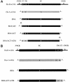
FIG. 1.
FIG. 1.
NA and HA genes used to generate recombinant influenza A/WSN viruses. (A) Schematic diagram of the influenza B/Yamagata/16/88 and A/WSN/33 parental and chimeric NA genes showing the ORFs of the NAs. NCRs are indicated by lines. CT, cytoplasmic tail; EC, ectodomain; TM, transmembrane domain. In BNA/A65 and BNA/ACT-ATM the NCRs have been extended by insertion of 65 nt into the 3′ NCR (nt 20 to 84) and insertion of 36 nt (nt 1298 to 1333) into the 5′ NCR, respectively. Flu B wt NA, influenza B/Yamagata wild-type virus NA; Flu A wt NA, influenza A/WSN wild-type virus NA. The asterisk in Flu B wild-type NA indicates the position of the start codon of the NB protein which is upstream of that of the NA protein. This upstream sequence coding for the amino terminus of the NB protein is not included in the chimeric constructs. (B) Schematic representation of the influenza B/Yamagata/16/88 and A/WSN/33 virus HA and the chimeric BHA-ACT/ATM segments. Flu B wt HA, influenza B/Yamagata wild-type virus HA; Flu A wt HA, influenza A/WSN wild-type virus HA. The ORFs are shown as boxes. The NCRs are represented as lines. CT, cytoplasmic tail; EC, ectodomain; TM, transmembrane domain. For details, see Materials and Methods.
FIG. 2.
FIG. 2.
Immunofluorescence analysis of MDCK cells infected with recombinant viruses. (A) Recombinant influenza A viruses expressing recombinant influenza B virus NAs. Subpanels: A and D, wild-type influenza B/Yamagata virus, B and C, wild-type influenza A/WSN virus; E and F, WSN-BNA virus; G and H, WSN-BNA/A65 virus; I and J, WSN-BNA/ACT virus; K and L, WSN-BNA/ACT-ATM virus. (B) Recombinant influenza A virus expressing the HA ectodomain of influenza B virus. Subpanels: A and B, mock infection; C and D WSN-BHA/ACT-ATM virus. α Flu B NA and α Flu B HA, monoclonal antibodies recognizing influenza B/Yamagata virus NA (9A6) or HA (15B6), respectively; α Flu A, polyclonal rabbit antiserum (i.e., 8236) raised against A/WSN virus.
FIG. 2.
FIG. 2.
Immunofluorescence analysis of MDCK cells infected with recombinant viruses. (A) Recombinant influenza A viruses expressing recombinant influenza B virus NAs. Subpanels: A and D, wild-type influenza B/Yamagata virus, B and C, wild-type influenza A/WSN virus; E and F, WSN-BNA virus; G and H, WSN-BNA/A65 virus; I and J, WSN-BNA/ACT virus; K and L, WSN-BNA/ACT-ATM virus. (B) Recombinant influenza A virus expressing the HA ectodomain of influenza B virus. Subpanels: A and B, mock infection; C and D WSN-BHA/ACT-ATM virus. α Flu B NA and α Flu B HA, monoclonal antibodies recognizing influenza B/Yamagata virus NA (9A6) or HA (15B6), respectively; α Flu A, polyclonal rabbit antiserum (i.e., 8236) raised against A/WSN virus.
FIG. 3.
FIG. 3.
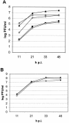
Growth characteristics of recombinant influenza viruses. MDCK cells were infected at an MOI of 0.001 with rWSN (⋄), WSN-BNA (▴), WSN-BNA/A65 (▵), WSN-BNA/ACT (▪), and WSN-BNA/ACT-ATM (□) viruses (A) or with wild-type WSN (♦) and WSN-BHA/ACT-ATM (P6 [▪] and H545Y [□]) viruses (B). Plaque assays were performed on MDCK cells. h p.i., hours postinfection.
FIG. 4.
FIG. 4.
Protein gel analysis of influenza A/WSN and recombinant WSN-BHA/ACT-ATM (P6) viruses. Proteins of purified viruses, either untreated (−) or treated with PNGase F (+), were separated by SDS-PAGE on a 12% gel and stained with Coomassie brilliant blue. Positions of untreated (−) and treated (+) proteins are indicated. The band of added PNGase F is shown to the right by an arrow. The positions of the deglycosylated influenza A/WSN HA1 (Flu A HA1) and WSN-BHA/ACT-ATM (Flu B HA1) viruses are indicated by arrows on the right. HA0, HA precursor protein; HA2, HA2 HA subunit.
FIG. 5.
FIG. 5.
Content of influenza B virus NA in purified chimeric viruses as determined by ELISA. ELISA plates were coated with 50 μl of serial twofold dilutions of recombinant purified WSN-BNA (▴), WSN-BNA/A65 (▵), WSN-BNA/ACT (▪), WSN-BNA/ACT-ATM (□), and wild-type B/Yamagata viruses (⋄). The starting concentration was 5 μg/ml. Plates were incubated with an influenza B virus NA-specific monoclonal antibody (9A6) and developed with a secondary horseradish peroxidase-conjugated anti-mouse IgG.

Similar articles

Cited by

References

    1. Baigent, S. J., and J. W. McCauley. 2001. Glycosylation of haemagglutinin and stalk-length of neuraminidase combine to regulate the growth of avian influenza viruses in tissue culture. Virus Res. 79:177-185. - PubMed
    1. Bilsel, P., M. R. Castrucci, and Y. Kawaoka. 1993. Mutations in the cytoplasmic tail of influenza A virus neuraminidase affect incorporation into virions. J. Virol. 67:6762-6767. - PMC - PubMed
    1. Crescenzo-Chaigne, B., N. Naffakh, and S. van der Werf. 1999. Comparative analysis of the ability of the polymerase complexes of influenza viruses types A, B, and C to assemble into functional RNPs that allow expression and replication of heterotypic model RNA templates in vivo. Virology 265:342-353. - PubMed
    1. Fodor, E., L. Devenish, O. G. Engelhardt, P. Palese, G. G. Brownlee, and A. García-Sastre. 1999. Rescue of influenza A virus from recombinant DNA. J. Virol. 73:9679-9682. - PMC - PubMed
    1. Fujii, Y., H. Goto, T. Watanabe, T. Yoshida, and Y. Kawaoka. 2003. Selective incorporation of influenza virus RNA segments into virions. Proc. Natl. Acad. Sci. USA 100:2002-2007. - PMC - PubMed

Publication types

MeSH terms

Substances

LinkOut - more resources