. 2003 Sep;77(17):9124-35.
doi: 10.1128/jvi.77.17.9124-9135.2003.

Targeting of the turnip yellow mosaic virus 66K replication protein to the chloroplast envelope is mediated by the 140K protein

Affiliations

Targeting of the turnip yellow mosaic virus 66K replication protein to the chloroplast envelope is mediated by the 140K protein

Delphine Prod'homme et al. J Virol. 2003 Sep.

Abstract

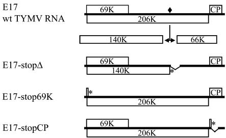
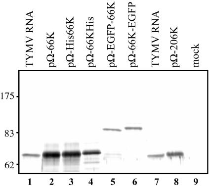
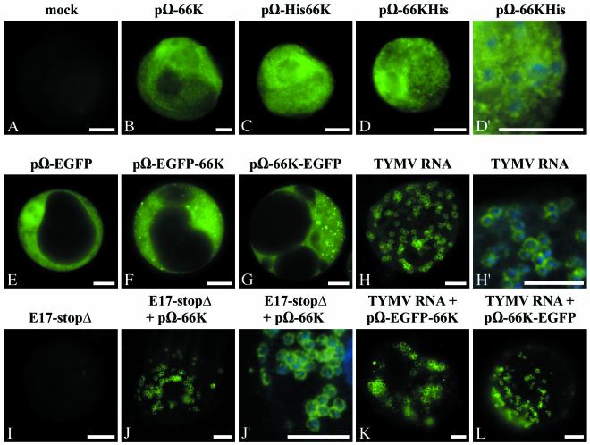
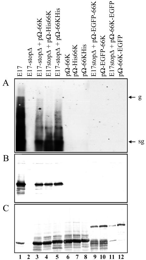
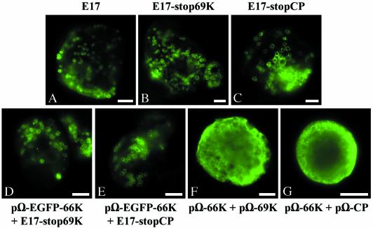
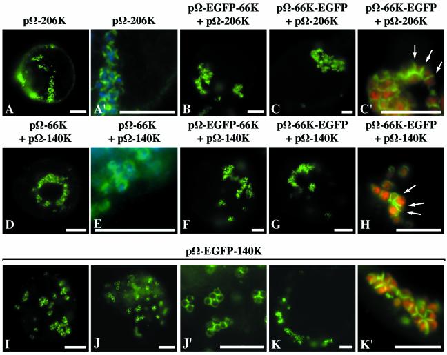
Turnip yellow mosaic virus (TYMV), a positive-strand RNA virus in the alphavirus-like superfamily, encodes two replication proteins, 140K and 66K, both being required for its RNA genome replication. The 140K protein contains domains indicative of methyltransferase, proteinase, and NTPase/helicase, and the 66K protein encompasses the RNA-dependent RNA polymerase domain. During viral infection, the 66K protein localizes to virus-induced chloroplastic membrane vesicles, which are closely associated with TYMV RNA replication. To investigate the determinants of its subcellular localization, the 66K protein was expressed in plant protoplasts from separate plasmids. Green fluorescent protein (GFP) fusion and immunofluorescence experiments demonstrated that the 66K protein displayed a cytoplasmic distribution when expressed individually but that it was relocated to the chloroplast periphery under conditions in which viral replication occurred. The 66K protein produced from an expression vector was functional in viral replication since it could transcomplement a defective replication template. Targeting of the 66K protein to the chloroplast envelope in the course of the viral infection appeared to be solely dependent on the expression of the 140K protein. Analysis of the subcellular localization of the 140K protein fused to GFP demonstrated that it is targeted to the chloroplast envelope in the absence of other viral factors and that it induces the clumping of the chloroplasts, one of the typical cytological effects of TYMV infection. These results suggests that the 140K protein is a key organizer of the assembly of the TYMV replication complexes and a major determinant for their chloroplastic localization and retention.


Figures

FIG. 1.
FIG. 1.
Schematic representation of the genomic organization of wild-type TYMV RNA and mutant genomes used in the present study. Open bars denote viral ORFs. The encoded 206K protein is proteolytically processed at a peptide bond signified by a filled square. Deletions are indicated by broken bars, and introduced stop codons are indicated by asterisks.
FIG. 2.
FIG. 2.
Expression of the TYMV 66K protein and its derivatives in plant cells. Arabidopsis protoplasts were transfected with TYMV RNA (lanes 1 and 7) or plasmids pΩ-66K (lane 2), pΩ-His66K (lane 3), pΩ-66KHis (lane 4), pΩ-EGFP-66K (lane 5), pΩ-66K-EGFP (lane 6), pΩ-206K (lane 8), and water (lane 9). The cells were harvested at 27 h posttransfection, and total proteins were extracted and subjected to SDS-8% PAGE and immunoblot analysis with anti-66K polyclonal antibodies. The position of molecular weight markers (Biolabs) is indicated.
FIG. 3.
FIG. 3.
The 66K protein and its derivatives have a cytoplasmic distribution when expressed individually but relocalize to the chloroplast envelope in infected cells. Arabidopsis protoplasts were transfected with water (A); with the expression plasmids pΩ-66K (B), pΩ-His66K (C), pΩ-66KHis (D and D′), pΩ-EGFP (E), pΩ-EGFP-66K (F), or pΩ-66K-EGFP (G); and with the viral RNA (H and H′). Transfections were also performed with transcript E17-stopΔ alone (I), or together with the plasmid pΩ-66K (J and J′), or with plasmid pΩ-EGFP-66K (K) or pΩ-66K-EGFP (L), together with viral RNA. The protoplasts were collected at 22 h posttransfection (H) or 46 h posttransfection (A to G and I to L), and they were either observed directly for GFP fluorescence (E to G, K, and L) or processed for indirect immunofluorescence labeling by using anti-66K antiserum followed by secondary antibodies coupled to Alexafluor-488 (A to D′ and H to J′). The cells were observed by epifluorescence microscopy. Scale bars, 10 μm. Panels D′, H′ and J′ are enlargements of panels D, H, and J, respectively, in which the DAPI staining (blue) of the chloroplastic DNA was acquired and superimposed to the fluorescence signal of the viral protein (green) to visualize the location of chloroplasts.
FIG. 4.
FIG. 4.
The 66K protein and some of its derivatives can support TYMV RNA replication in trans. Arabidopsis protoplasts were transfected with transcript E17 (lane 1), transcript E17-stopΔ (lane 2), or combinations of transcript E17-stopΔ with the plasmids pΩ-66K (lane 3), pΩ-His66K (lane 4), pΩ-66KHis (lane 5), pΩ-EGFP-66K (lane 9), and pΩ-66K-EGFP (lane 11). Each of the expression plasmids was also transfected individually (lanes 6 to 8 and lanes 10 and 12, respectively). The transfected protoplasts were collected at 46 h posttransfection and used both for RNA and protein analyses. (A) Equivalent amounts of total RNA prepared from the transfected protoplasts were analyzed by Northern blotting with a single-stranded, DIG-labeled RNA probe complementary to positive-strand TYMV RNA. The migration positions of genomic (g) and subgenomic (sg) RNAs are indicated. (B and C) Equivalent amounts of proteins were analyzed by SDS-PAGE on 15% (B) or 8% (C) polyacrylamide gels. The gels were electroblotted onto a nitrocellulose filter, and proteins were revealed by Western blotting with anti-CP (B) or anti-66K (C) antiserum.
FIG. 5.
FIG. 5.
Targeting of the 66K protein to the chloroplast envelope is not dependent on the 69K movement protein or CP. Arabidopsis protoplasts were transfected with transcript E17 (A), with transcript E17-stop69K alone (B) or together with plasmid pΩ-EGFP-66K (D), with transcript E17-stopCP alone (C) or together with plasmid pΩ-EGFP-66K (E), or with plasmid pΩ-66K together with plasmid pΩ-69K (F) or pΩ-CP (G). The protoplasts were collected at 25 h (A to C) or 46 h (D to G) posttransfection, and they were either observed directly for GFP fluorescence (D to E) or they were processed for indirect immunofluorescence labeling by using anti-66K antiserum, followed by secondary antibodies coupled to Alexafluor-488 (A to C and F to G). The cells were observed by epifluorescence microscopy. Scale bars, 10 μm.
FIG. 6.
FIG. 6.
Targeting of the 66K protein to the chloroplast envelope is dependent on the 140K protein, which localizes to the chloroplast envelope in the absence of other viral components. Arabidopsis protoplasts were transfected with the plasmid pΩ-206K alone (A and A′) or pΩ-206K together with the plasmids pΩ-EGFP-66K (B) or pΩ-66K-EGFP (C and C′) or with the plasmid pΩ-140K together with the plasmids pΩ-66K (D and E), pΩ-EGFP-66K (F), or pΩ-66K-EGFP (G and H), or with the plasmid pΩ-EGFP-140K (I to K′). The protoplasts were collected at 22 h (A and A′) or 46 h (B to K′) posttransfection, and they were either observed directly for GFP fluorescence (B to C′ and F to K′) or they were processed for indirect immunofluorescence labeling by using anti-66K antiserum, followed by secondary antibodies coupled to Alexafluor-488 (A, A′, D, and E). The cells were observed by epifluorescence microscopy. Scale bars, 10 μm. Panels A′, C′, J′, and K′ are enlargements of panels A, C, J, and K, respectively. In order to visualize the location of chloroplasts, the DAPI staining (blue) of the chloroplastic DNA was acquired and superimposed onto the fluorescence signal of the viral protein (green) in panels A′ and E, whereas in panels C′, H, and K′ the chlorophyll autofluorescence (red) was acquired and superimposed onto the GFP fluorescence (green).

Similar articles

Cited by

References

    1. Ahola, T., A. Lampio, P. Auvinen, and L. Kaariainen. 1999. Semliki Forest virus mRNA capping enzyme requires association with anionic membrane phospholipids for activity. EMBO J. 18:3164-3172. - PMC - PubMed
    1. Ausubel, F. M., R. Brent, R. E. Kingston, D. D. Moore, J. G. Seidman, J. A. Smith, and K. Struhl. 1987. Current protocols in molecular biology. Wiley, New York, N.Y.
    1. Barends, S., H. H. Bink, S. H. van den Worm, C. W. Pleij, and B. Kraal. 2003. Entrapping ribosomes for viral translation: tRNA mimicry as a molecular Trojan horse. Cell 112:123-129. - PubMed
    1. Bass, B. L. 2000. Double-stranded RNA as a template for gene silencing. Cell 101:235-238. - PubMed
    1. Bransom, K. L., S. E. Wallace, and T. W. Dreher. 1996. Identification of the cleavage site recognized by the turnip yellow mosaic virus protease. Virology 217:404-406. - PubMed

Publication types

MeSH terms

LinkOut - more resources