Comparative Study
. 2003 Sep;77(17):9136-46.
doi: 10.1128/jvi.77.17.9136-9146.2003.

A wild-type porcine encephalomyocarditis virus containing a short poly(C) tract is pathogenic to mice, pigs, and cynomolgus macaques

Affiliations
Comparative Study

A wild-type porcine encephalomyocarditis virus containing a short poly(C) tract is pathogenic to mice, pigs, and cynomolgus macaques

Rebecca LaRue et al. J Virol. 2003 Sep.

Abstract

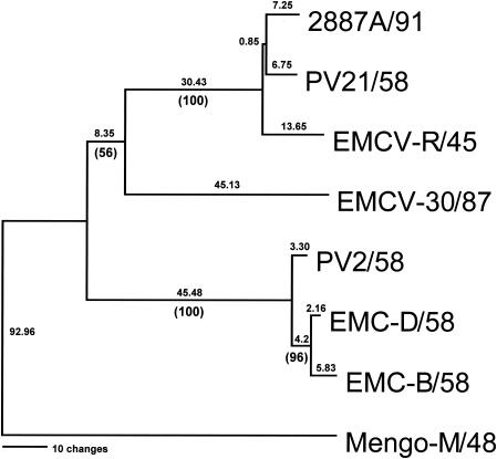
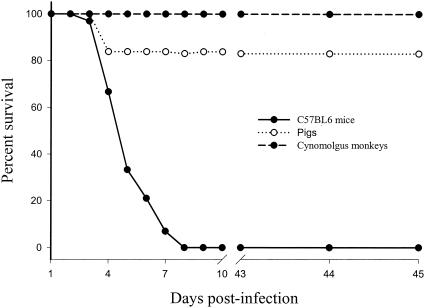
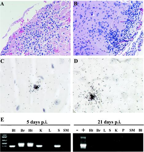
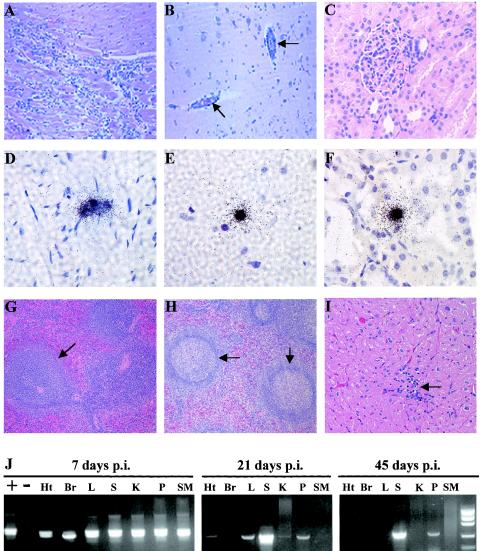
Previous studies using wild-type Encephalomyocarditis virus (EMCV) and Mengo virus, which have long poly(C) tracts (61 to 146 C's) at the 5' nontranslated region of the genome, and variants of these viruses genetically engineered to truncate or substitute the poly(C) tracts have produced conflicting data on the role of the poly(C) tract in the virulence of these viruses. Analysis of the nucleotide sequence of an EMCV strain isolated from an aborted swine fetus (EMCV 30/87) revealed that the virus had a poly(C) tract that was 7- to 10-fold shorter than the poly(C) tracts of other EMCV strains and 4-fold shorter than that of Mengo virus. Subsequently, we investigated the virulence and pathogenesis of this naturally occurring short-poly(C)-tract-containing virus in rodents, pigs, and nonhuman primates. Infection of C57BL/6 mice, pigs, and cynomolgus macaques resulted in similar EMCV 30/87 pathogenesis, with the heart and brain as the primary sites of infections in all three animals, but with different disease phenotypes. Sixteen percent of EMCV 30/87-infected pigs developed acute fatal cardiac failure, whereas the rest of the pigs were overtly asymptomatic for as long as 90 days postinfection (p.i.), despite extensive myocardial and central nervous system (CNS) pathological changes. In contrast, mice infected with >/==" BORDER="0">4 PFU of EMCV 30/87 developed acute encephalitis that resulted in the death of all animals (n = 25) between days 2 and 7 p.i. EMCV 30/87-infected macaques remained overtly asymptomatic for 45 days, despite extensive myocardial and CNS pathological changes and viral persistence in more than 50% of the animals. The short poly(C) tract in EMCV 30/87 (CUC(5)UC(8)) was comparable to that of strain 2887A/91 (C(10)UCUC(3)UC(10)), another recent porcine isolate.


Figures

FIG. 1.
FIG. 1.
Phylogenetic relationship between EMCV 30/87 and other EMCV strains. Following alignment of the contiguous predicted amino acid sequences for the translated regions of the viruses (LP, VP4, VP2, VP3, VP1, 2A, 2B, 2C, 3A, 3B, 3C, and 3D), a rooted phylogram was generated by maximum parsimony analysis using Mengo virus M/48 as the outgroup. Absolute distances are listed above each branch, with bootstrap confidence levels given below in parentheses. GenBank accession numbers for the viruses are given in Materials and Methods.
FIG. 2.
FIG. 2.
Survival curves of EMCV-infected mice, pigs, and macaques. Six-week-old C57BL/6 mice (n = 30), 5-week-old pigs (n = 35), and 2- to 4-year-old cynomolgus macaques (n = 12) were intraperitoneally inoculated with EMCV 30/87 and monitored daily for clinical signs and fatality for 45 days. Sham-inoculated pigs (n = 2), mice (n = 4), and macaques (n = 2) remained asymptomatic and had no fatalities.
FIG. 3.
FIG. 3.
Pathological changes and detection of viral RNA in mice infected with EMCV 30/87. Five-week-old C57BL/6 mice were intraperitoneally inoculated with 102 to 103 PFU of EMCV 30/87, and histopathologic changes and the presence of EMCV RNA in the blood (Bl), heart (Ht), brain (Br), spleen (S), pancreas (P), liver (L), kidney (K), and skeletal muscle (SM) were analyzed at 2 to 8 days p.i. All EMCV 30/87-infected mice inoculated with more than 102 PFU of virus died between days 2 and 8 p.i. Histopathologic changes were confined to the brain and heart. In the heart, there were large foci of lymphocytic infiltration in the myocardium (A), accompanied by degeneration and necrosis of cardiac myocytes, whereas in the brain, similar lymphocytic infiltrations were observed in the cerebral cortices (B), hypothalami, and hippocampi. EMCV RNA was detected in both the heart (C) and brain (D) by in situ hybridization, but also in the blood, kidney, spleen, and skeletal muscle by RT-PCR (E). In the brain and heart, which had high levels of EMCV RNA, nested RT-PCR using VP2 primers followed by electrophoresis resulted in two visible bands, the larger (499 bp) representing the primary reaction product and the smaller (350 bp) representing the nested reaction product. For histopathology, heart and brain sections were embedded in paraffin and 4-μm-thick sections were stained with hematoxylin and eosin. In situ hybridization was performed using a 35S-labeled VP1 cDNA probe. Results for mice inoculated with 2 to 10 PFU are shown in the right portion of panel E.
FIG. 4.
FIG. 4.
EMCV-induced pathological changes and detection of viral RNA in pigs. Five-week-old pigs were intraperitoneally inoculated with 2.9 × 10⁸ PFU of EMCV 30, and histopathologic changes and the presence of EMCV RNA in the heart, brain, spleen, pancreas, liver, kidney, and skeletal muscle were analyzed 7, 21, 45, and 90 days p.i. In the acute phase (7 days p.i.), inflammation and degenerative changes were observed in the heart (A), and brain (B), but no changes were observed in the other organs. In the chronic phase (21 to 90 days p.i), infiltration of lymphocytes in the heart (C) and perivascular cuffing in the brain (D) were evident throughout the infection period up to day 90 p.i. and were accompanied by persistence, as demonstrated by localization of EMCV RNA by in situ hybridization (E and F). For histopathology, heart and brain sections were embedded in paraffin, and 4-μm-thick sections were stained with hematoxylin and eosin. In situ hybridization was performed by using a 35S-labeled VP1 cDNA probe.
FIG.5.
FIG.5.
EMCV-induced pathological changes and detection of viral RNA in cynomolgus macaques. Adult macaques were intraperitoneally inoculated with 2.9 × 10⁸ PFU of EMCV 30/87, and histopathologic changes were analyzed 7, 21, and 45 days later. At day 7 p.i., there were acute inflammatory and mild degenerative changes in the myocardium (A) and brain (B, arrows), whereas there were no detectable changes in kidneys (C), livers, and spleens from the same animals. In contrast, EMCV RNA was widely distributed in tissues at day 7 p.i., as demonstrated by in situ hybridization (black grains) in the myocardium (D), brain (E), and kidneys (F) and by RT-PCR in the heart (Ht), brain (Br), liver (L), spleen (S), kidney (K), pancreas (P), and skeletal muscle (SM) (J). At days 21 and 45 p.i., mild lymphocytic infiltration was observed in the heart (I, arrow), and the formation of prominent germinal centers was observed in the spleen (H, arrows), in contrast to spleens from 7-day EMCV-infected macaques (G, arrow). EMCV RNA was most consistently observed in the spleen and pancreas at days 21 and 45 p.i. (J). In tissues with high levels of EMCV RNA at days 7 (all tissues), 21 (spleen), and 45 (spleen) p.i., nested RT-PCR using the VP2 primers followed by agarose gel electrophoresis resulted in two visible bands, the larger (499 bp) representing the primary reaction product and the smaller (350 bp) representing the nesting reaction product. For histopathology, macaque tissue sections were embedded in paraffin and 4-μm-thick sections were stained with hematoxylin and eosin. In situ hybridization was performed using a 35S-labeled VP1 cDNA probe.
FIG. 6.
FIG. 6.
Detection by ELISA of EMCV-specific IgG in serum samples from experimentally infected cynomolgus macaques. (A) Virus-specific IgG levels in serum samples from macaques infected for 7, 21, or 45 days. Data are presented as mean A630 readings (± standard errors) from four serum samples at each time point performed at serum dilutions between 1:500 and 1:2,000. (B) Profile of virus-specific IgG levels through the 45-day experimental period as determined at a 1:500 serum dilution.

Similar articles

Cited by

References

    1. Bae, Y. S., H. M. Eun, and J. W. Yoon. 1989. Genomic differences between diabetogenic and nondiabetogenic variants of encephalomyocarditis virus. Virology 170:282-287. - PubMed
    1. Blanchard, J. L., K. F. Soike, and G. B. Baskin. 1987. Encephalomyocarditis virus infection in African green and squirrel monkeys: comparison of pathological effects. Lab. Anim. Sci. 37:635-639. - PubMed
    1. Brewer, L. A., C. Brown, M. P. Murtaugh, and M. K. Njenga. Transmission of porcine encephalomyocarditis virus (EMCV) to mice by transplanting EMCV-infected pig tissues. Xenotransplantation, in press. - PubMed
    1. Brewer, L. A., H. C. M. Lwamba, M. P. Murtaugh, A. C. Palmenberg, C. Brown, and M. K. Njenga. 2001. Porcine encephalomyocarditis virus persists in pig myocardium and infects human myocardial cells. J. Virol. 75:11621-11629. - PMC - PubMed
    1. Cerutis, D. R., R. H. Bruner, D. C. Thomas, and D. J. Giro. 1989. Tropism and histopathology of the D, B, K and MM variants of encephalomyocarditis virus. J. Med. Virol. 29:63-69. - PubMed

Publication types

MeSH terms

LinkOut - more resources