. 2003 Sep;77(17):9173-82.
doi: 10.1128/jvi.77.17.9173-9182.2003.

Tsg101 control of human immunodeficiency virus type 1 Gag trafficking and release

Affiliations

Tsg101 control of human immunodeficiency virus type 1 Gag trafficking and release

A Goff et al. J Virol. 2003 Sep.

Abstract

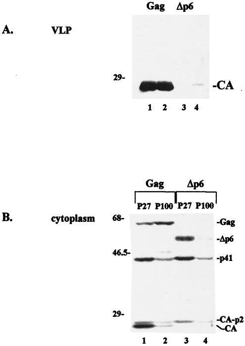
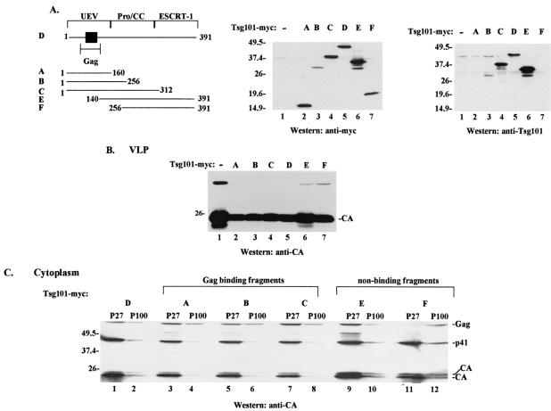
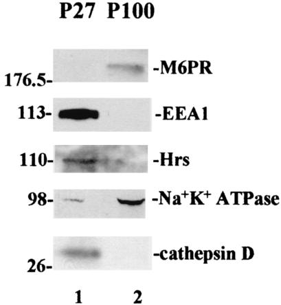
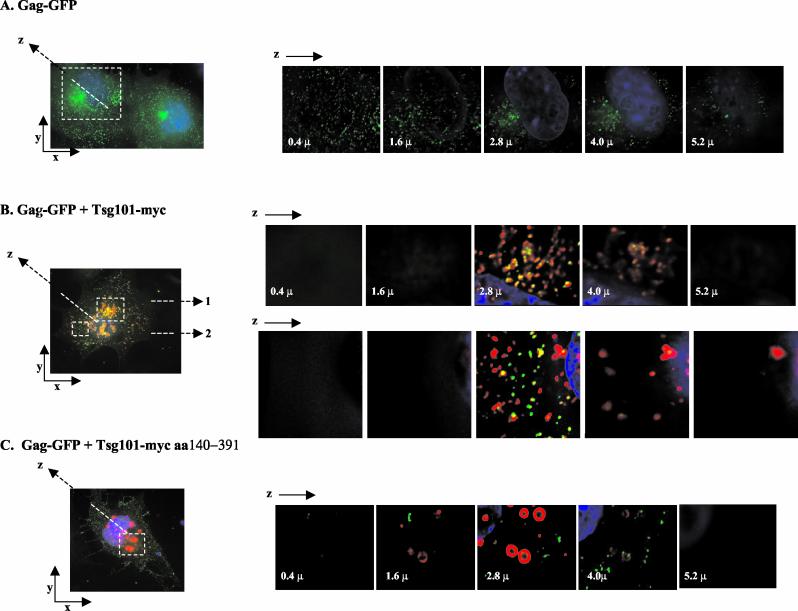
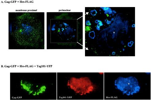
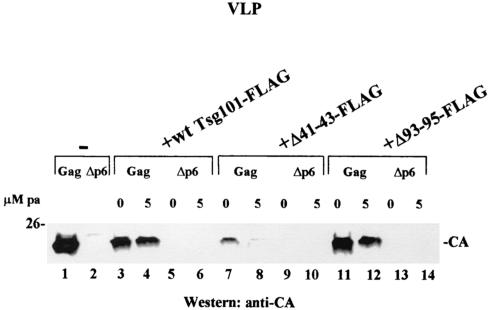
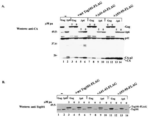
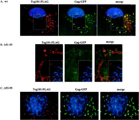
The structural precursor polyprotein of human immunodeficiency virus type 1, Pr55(gag), contains a proline-rich motif (PTAP) called the "late domain" in its C-terminal p6 region that directs release of mature virus-like particles (VLPs) from the plasma membranes of gag-transfected COS-1 cells. The motif binds Tsg101 (vacuolar protein-sorting protein 23, or Vps23), which functions in endocytic trafficking. Here, we show that accumulation of the wild-type (wt) Gag precursor in a fraction of COS-1 cytoplasm enriched in multivesicular bodies and small particulate components of the plasma membrane (P100) is p6 dependent. Cleavage intermediates and mature CA mainly partitioned with more rapidly sedimenting larger material enriched in components of lysosomes and early endosomes (P27), and this also was p6 dependent. Expression of truncated or full-length Tsg101 proteins interfered with VLP assembly and Gag accumulation in the P100 fraction. This correlated with reduced accumulation of Gag tagged with green fluorescent protein (Gag-GFP) at the plasma membrane and colocalization with the tagged Tsg101 in perinuclear early endosomes, as visualized by confocal microscopy. Fractionation analysis and confocal examination both indicated that the N-terminal region of Tsg101, which contains binding sites for PTAP and ubiquitin (Ub), was required for Gag trafficking to the plasma membrane. Expression of FLAG-tagged Tsg101 with a deletion in the Ub-binding pocket inhibited VLP release almost completely and to a significantly greater extent than expression of the wt tagged Tsg101 protein or Tsg101-FLAG containing a deletion in the PTAP-binding region. The results demonstrate that Gag associates with endosomal trafficking compartments and indicate that efficient release of virus particles from the plasma membrane requires both the PTAP- and Ub-binding functions of Tsg101 to recruit the cellular machinery required for budding.


Figures

FIG. 1.
FIG. 1.
p6 dependence of VLP release and intracellular Gag localization. VLPs (A) and cytoplasmic extracts (B) were prepared from cells transfected with wt gag or gag-pol (lanes 1 and 2) or Δp6 gag or gag-pol (lanes 3 and 4). Duplicate VLP samples are shown. Cytoplasmic extracts derived from cells expressing wt (lanes 1 and 2) or Δp6 (lanes 3 and 4) Gag were fractionated sequentially to obtain the P27 (lanes 1 and 3) and P100 (lanes 2 and 4) fractions. Proteins were identified by Western blotting with an anti-CA antibody. Molecular weight markers (in thousands) are given on the left.
FIG. 2.
FIG. 2.
Effects of Tsg101 overexpression on VLP release and Gag accumulation in particulate cytoplasmic fractions. (A) (Left) Schematic diagram showing full-length and truncated Tsg101 proteins used in this study, as follows: fragment A, aa 1 to 160; fragment B, aa 1 to 256; fragment C, aa 1 to 312; Tsg101 protein D, aa 1 to 391; fragment E, aa 140 to 391; fragment F, aa 256 to 391. Filled square, Gag binding region in the UEV domain. (Center) Western blot analysis of cytoplasmic extracts for adventitious Tsg101 expression by use of an anti-Myc antibody. (Right) The blot was reprobed with an anti-Tsg101 monoclonal antibody. The antibody recognizes full-length Tsg101 and fragments B, C, and E. Molecular weight markers are shown to the left of the gels. (B) VLPs were isolated from the culture media of cells transfected with DNA encoding Gag or Gag-Pol and the empty vector (lane 1), full-length Tsg101-Myc (lane 5), or fragments of Tsg101-Myc (lanes 2 to 4, 6, and 7). (C) P27 (lanes 1, 3, 5, 7, 9, and 11) and P100 (lanes 2, 4, 6, 8, 10, and 12) fractions were isolated from cytoplasmic extracts prepared from the cells.
FIG. 3.
FIG. 3.
Examination of P27 and P100 fractions for endogenous markers of endosomal compartments. P27 (lane 1) and P100 (lane 2) fractions prepared from cells expressing Gag were examined for endosomal protein markers by Western blotting using marker-specific antibodies.
FIG. 4.
FIG. 4.
Confocal microscopy of cells expressing Gag-GFP and wt or N-terminally truncated Tsg101-Myc. Shown are confocal images of fixed COS-1 cells transfected with Gag-GFP alone (A), Gag-GFP and wt Tsg101-Myc (B), or Gag-GFP and N-terminally truncated Tsg101-Myc (fragment E) (C). Serial z sections from the adherent cell surface through the nucleus are shown for the region indicated by the dashed white square. In panel B, view 1 corresponds to the upper white square and view 2 corresponds to the lower white square. Green, Gag; red, Tsg101-Myc; blue, nuclei stained with DAPI.
FIG. 5.
FIG. 5.
Confocal microscopy of cells expressing Gag-GFP, Hrs-FLAG, and Tsg101-YFP. Shown are confocal images of fixed COS-1 cells transfected with Gag-GFP and Hrs-FLAG (A) or Gag-GFP, Hrs-FLAG, and Tsg101-YFP (B). Cells expressing Gag-GFP and Hrs-FLAG contained fluorescence in peripheral and perinuclear regions, and both regions are shown. Fluorescence was exclusively perinuclear in cells expressing Gag-GFP, Hrs-FLAG, and Tsg101-YFP. The area within the dashed white square in panel A was deconvolved and enlarged to enhance visualization of Hrs-Gag colocalization. Green, Gag; red, Tsg101-YFP; blue, Hrs-FLAG. There is some nonspecific staining of nuclei (n) with the anti-FLAG antibody.
FIG. 6.
FIG. 6.
Effects of N-terminal Tsg101 mutations on VLP release. VLPs were isolated from the media of cells expressing wt Gag (lanes 1, 3, 4, 7, 8, 11, and 12) or the Δp6 precursor protein (lanes 2, 5, 6, 9, 10, 13, and 14).
FIG. 7.
FIG. 7.
Effects of N-terminal Tsg101 mutations on Gag and Tsg101 accumulation in the cytoplasm. Cytoplasmic extracts were prepared from cells expressing Gag (lanes 1, 3, 4, 7, 8, 11, and 12) or the Δp6 protein precursor (lanes 2, 5, 6, 9, 10, 13, and 14) and either wt Tsg101-FLAG or the indicated Tsg101-FLAG mutant and were analyzed by Western blotting using an anti-CA (A) or anti-Tsg101 (B) antibody.
FIG. 8.
FIG. 8.
Effects of N-terminal Tsg101 mutations on Gag and Tsg101 distribution. Shown are confocal images of COS-1 cells transfected with Gag-GFP and either wt Tsg101-FLAG (A), Δ41-43 Tsg101-FLAG (B), or Δ93-95 Tsg101-FLAG (C) in the absence of pa. Red fluorescence indicates leaky (uninduced) Tsg101-FLAG expression from the transfected tsg101 DNA. Areas within dashed white rectangles were deconvolved to enhance the resolution. Green, Gag; red, Tsg101-FLAG; yellow, colocalization.

Similar articles

Cited by

References

    1. Alland, L., S. M. Peseckis, R. E. Atherton, L. Berthiaume, and M. D. Resh. 1994. Dual myristylation and palmitylation of Src family member p59fyn affects subcellular localization. J. Biol. Chem. 269:16701-16705. - PubMed
    1. Babst, M., D. J. Katzmann, E. J. Estepa-Sabal, T. Meerloo, and S. D. Emr. 2002. ESCRT-III: an endosome-associated heterooligomeric protein complex required for MVB sorting. Dev. Cell 3:271-282. - PubMed
    1. Babst, M., D. J. Katzmann, W. B. Synder, B. Wendland, and S. D. Emr. 2002. Endosome-associated complex, ESCRT-II, recruits transport machinery for protein sorting at the multivesicular body. Dev. Cell 3:282-289. - PubMed
    1. Babst, M., G. Odorizzi, E. J. Estepa, and S. D. Emr. 2000. Mammalian tumor susceptibility gene 101 (Tsg101) and the yeast homologue, Vps23p, both function in late endosomal trafficking. Traffic 1:248-258. - PubMed
    1. Bishop, N., A. Horman, and P. Woodman. 2002. Mammalian class E vps proteins recognize ubiquitin and act in the removal of endosomal protein-ubiquitin conjugates. J. Cell Biol. 57:91-101. - PMC - PubMed

Publication types

MeSH terms