. 2003 Sep;77(17):9192-203.
doi: 10.1128/jvi.77.17.9192-9203.2003.

Association of the herpes simplex virus type 1 Us11 gene product with the cellular kinesin light-chain-related protein PAT1 results in the redistribution of both polypeptides

Affiliations

Association of the herpes simplex virus type 1 Us11 gene product with the cellular kinesin light-chain-related protein PAT1 results in the redistribution of both polypeptides

Louisa Benboudjema et al. J Virol. 2003 Sep.

Abstract

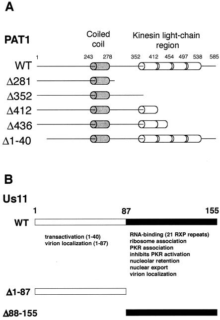
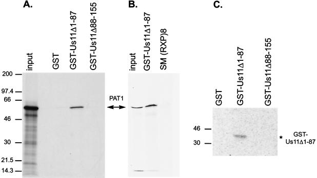
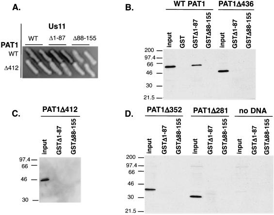
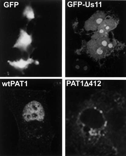
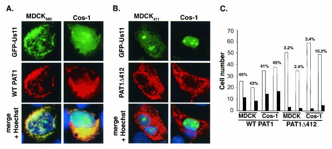
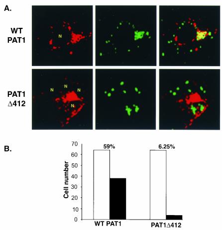
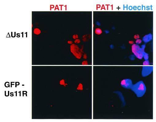
The herpes simplex virus type 1 (HSV-1) Us11 gene encodes a multifunctional double-stranded RNA (dsRNA)-binding protein that is expressed late in infection and packaged into the tegument layer of the virus particle. As a tegument component, Us11 associates with nascent capsids after its synthesis late in the infectious cycle and is delivered into newly infected cells at times prior to the expression of viral genes. Us11 is also an abundant late protein that regulates translation through its association with host components and contains overlapping nucleolar retention and nuclear export signals, allowing its accumulation in both nucleoli and the cytosol. Thus, at various times during the viral life cycle and in different intracellular compartments, Us11 has the potential to execute discrete tasks. The analysis of these functions, however, is complicated by the fact that Us11 is not essential for viral replication in cultured cells. To discover new host targets for the Us11 protein, we searched for cellular proteins that interact with Us11 and have identified PAT1 as a Us11-binding protein according to multiple, independent experimental criteria. PAT1 binds microtubules, participates in amyloid precursor protein trafficking, and has homology to the kinesin light chain (KLC) in its carboxyl terminus. The carboxyl-terminal dsRNA-binding domain of Us11, which also contains the nucleolar retention and nuclear export signals, binds PAT1, whereas 149 residues derived from the KLC homology region of PAT1 are important for binding to Us11. Both PAT1 and Us11 colocalize within a perinuclear area in transiently transfected and HSV-1-infected cells. The 149 amino acids derived from the KLC homology region are required for colocalization of the two polypeptides. Furthermore, although PAT1 normally accumulates in the nuclear compartment, Us11 expression results in the exclusion of PAT1 from the nucleus and its accumulation in the perinuclear space. Similarly, Us11 does not accumulate in the nucleoli of infected cells that overexpress PAT1. These results establish that Us11 and PAT1 can associate, resulting in an altered subcellular distribution of both polypeptides. The association between PAT1, a cellular trafficking protein with homology to KLC, and Us11, along with a recent report demonstrating an interaction between Us11 and the ubiquitous kinesin heavy chain (R. J. Diefenbach et al., J. Virol. 76:3282-3291, 2002), suggests that these associations may be important for the intracellular movement of viral components.


Figures

FIG. 1.
FIG. 1.
Summary of structural and functional motifs found in PAT1 and Us11. (A) Schematic representation of sequence motifs present in the WT PAT1 protein and the mutants used in the present study. The full-length PAT1 protein is composed of 585 amino acid residues, binds to microtubules, and is involved in the intracellular trafficking of APP. The protein contains a segment predicted to adopt the structure of a coiled coil (amino acids 243 to 278) and a region that shares homology with kinesin light chain (amino acids 352 to 538). Mutant PAT1 proteins used in the present study and the regions they contain are also illustrated. (B) Illustration of functional domains in the HSV-1 Us11 protein. The Us11 protein encoded by HSV-1 (Patton strain) is 155 amino acids long. The carboxyl-terminal 68-amino-acid domain contains a novel dsRNA-binding motif composed primarily of 21 repeats of the amino acid triplet RXP. The activities associated with the amino and carboxyl-terminal domains and a representation of the GST fusion proteins are shown.
FIG. 2.
FIG. 2.
PAT1 specifically interacts with the carboxyl-terminal 68 amino acids of Us11. (A) 35S-labeled PAT1 was incubated with purified GST fusion proteins containing the C-terminal domain of Us11 (Us11Δ1-87), the N-terminal domain of Us11 (Us11Δ88-155), or GST. After the complexes were isolated on glutathione-agarose beads, the proteins were separated by SDS-PAGE, and the fixed dried gel was exposed to Kodak film. The major translated product (input) corresponds to full-length PAT1. PAT1 specifically forms a complex with the C-terminal 87 amino acids of Us11. (B) GST fusion proteins were incubated with lysates prepared from 293T cells transfected with plasmids expressing FLAG-tagged, full-length PAT1. GST-containing complexes were collected on glutathione agarose beads, fractionated by electrophoresis in SDS-polyacrylamide gels, and transferred to a PVDF membrane. The membrane was probed with anti-FLAG antibody, followed by a horseradish peroxidase- conjugated secondary antibody, and PAT1 was visualized via enhanced chemiluminescence. (C) GST and the two GST-Us11 fusion proteins (GST-Us11Δ1-87 and GST-Δ88-155) were subjected to electrophoresis in SDS-polyacrylamide gels, blotted onto nitrocellulose membranes, renatured, and then probed with in vitro-translated [35S]methionine-labeled PAT1. The filter was washed and subsequently exposed in a phosphorimager cassette.
FIG. 3.
FIG. 3.
PAT1 residues 412 to 585 containing the kinesin light-chain homology region are required to interact with the C-terminal domain of Us11. (A) Full-length PAT1, PAT1 Δ281, Δ352, Δ412, Δ436, and Δ1-40 were expressed in yeast as activation domain fusions. WT Us11, Us11Δ1-87, and Us11Δ88-155 were expressed in yeast fused to the LexA DNA-binding domain. Interaction between Us11 and PAT1 was detected by the ability to form blue colonies on indicator plates. PAT1Δ412, which lacks amino acids 412 to 585 containing the kinesin light-chain repeats, does not interact with the C-terminal domain of Us11 and appears as a representative example. (B) Cell-free lysates were prepared from Cos cells transfected with plasmids expressing either FLAG-tagged WT PAT1 or PAT1 Δ281, Δ352, Δ412, or Δ436 (input). Equal amounts of purified GST, GST-Us11Δ1-87, or GST-US11Δ88-155 were incubated in the extracts. After the GST proteins were collected on glutathione-agarose beads, the complexes were then washed and fractionated by SDS-PAGE. The proteins were transferred to a solid support, reacted with antibodies, and visualized as described in the legend to Fig. 2.
FIG. 4.
FIG. 4.
Subcellular localization of GFP Us11, PAT1, or PAT1Δ412 after expression of each protein individually in transfected cells. MDCK cells were fixed and permeabilized 24 to 48 h after transfection with plasmids expressing either GFP, GFP-Us11, FLAG-tagged PAT1, or FLAG-tagged PAT1Δ412. They were subsequently examined for intrinsic GFP fluoresence or processed for immunofluorescence to detect PAT1.
FIG. 5.
FIG. 5.
Redistribution of PAT1 and colocalization of PAT1 and Us11 require 173 C-terminal residues of PAT1 that contain homology to KLC. MDCK585 (A) or MDCK411 (B) cells were transfected with 7 μg of pEGFP-Us11. Cos-1 cells were cotransfected with 7 μg of pEGFP-Us11 and an additional plasmid (7 μg) expressing either FLAG-tagged WT PAT1 (A) or PAT1Δ412 (B) polypeptides. At 24 to 48 h after transfection, the cells were processed for immunofluoresence to detect PAT1. Nuclei were stained with Hoechst stain. The distribution of GFP-Us11 was monitored by its intrinsic fluorescence. (C) Cells expressing both GFP-Us11 and either PAT1 or PAT1Δ412 were counted (open bars), and the numbers of cells in which Us11 and PAT1 colocalized were recorded (solid bars). The percentage of cells that expressed both GFP-Us11 and PAT1 (WT or Δ412) in which colocalization was observed appears on top of the bar graph for each experiment. Whereas on average, 44% of cells expressing both GFP-Us11 and PAT1 demonstrated colocalization of both proteins, only 5% of cells that expressed GFP-Us11 and PAT1Δ412 exhibited colocalization of the polypeptides.
FIG. 6.
FIG. 6.
Structure of ΔUs11 and GFP-Us11 recombinant viruses. (A) The HSV-1 genome is depicted with the unique long (UL), unique short (Us), and repetitive segments (rectangles) delineated. (B) Expanded view of the WT Us-TRs junction region is shown along with pertinent restriction enzyme cleavage sites. The Us10, Us11, and Us12 ORFs appear as boxes. cis-Acting promoter elements are indicated by stars, and each promoter is normally associated with either the Us10, Us11, or Us12 ORF (denoted by the numbers 10, 11, or 12 at the lower right of each star). Arrows located above each ORF represent mRNA transcripts produced from each promoter. All three transcripts are polyadenylated at a common site downstream from the 3′ end of the Us10 ORF. The noncontiguous segment of the Us12 transcript denotes an RNA splicing event. (C) An expanded view of the Us-TRs junction in the ΔUs11 recombinant virus at which the Us11 ORF has been deleted and replaced with the GFP ORF, followed by a heterologous polyadenylation signal. As a consequence of this mutation, the Us10 ORF is disrupted (the breakpoint is denoted by a broken line at the right end of the box), the Us10 promoter embedded in the Us11 ORF is deleted, and an mRNA capable of encoding the Us10 polypeptide is not produced. (D) An expanded view of the Us-TRs junction in the GFP-Us11R recombinant virus that expresses a GFP-Us11 fusion protein. The GFP ORF has been fused to the 5′ end of the Us11 ORF such that a GFP-Us11 fusion protein is produced. Note that the Us10 ORF and promoter are intact and a Us10 mRNA is produced.
FIG. 7.
FIG. 7.
Construction and characterization of a recombinant HSV-1 expressing an EGFP Us11 fusion protein. (A) Southern analysis of the Us-TRs junction fragments. Viral (GFP-Us11R, ΔUs11, and WT) or plasmid (pΔUs11) DNA was digested to completion with EcoNI, fractionated by electrophoresis in a 1% agarose gel, and transferred onto a nylon membrane. After the blot was probed with a 32P-labeled NcoI-PflMI DNA fragment derived from the Us-TRs region shown in Fig. 6B, the washed membrane was exposed to X-ray film. The plasmid pΔUs11 was used to construct the recombinant virus ΔUs11. EcoNI-digested viral DNA from two different isolates of ΔUs11 appears as a standard. The genetic structure that surrounds the Us-TRs junction in both the plasmid (pΔUs11) and the ΔUs11 viruses is depicted in Fig. 6C. ΔUs11 was the parental virus from which the GFP-Us11R recombinants were constructed. Two plaque-purified isolates that contain a GFP-Us11 fusion gene as depicted in Fig. 6D are shown. (B) Pattern of late protein synthesis in cells infected with a recombinant virus that expresses a GFP-Us11 fusion protein. Vero cells were infected with WT HSV-1, the ΔUs11 mutant, or the GFP-Us11R virus. At 16.5 h postinfection, the cultures were labeled with 35S-labeled amino acids for 1 h, the samples were solubilized in SDS-PAGE loading buffer, and the proteins were fractionated in SDS-polyacrylamide gels. The fixed, dried gel was subsequently exposed to X-ray film. Molecular mass standards (in kilodaltons) appear to the left of the panel. (C) The GFP-Us11R recombinant virus expresses a GFP-Us11 fusion protein. Total protein was isolated from cells infected with either WT, ΔUs11, or GFP-Us11R HSV-1. After fractionation by SDS-PAGE, the polypeptides were electrophoretically transferred to a membrane and incubated with either anti-Us11 or anti-GFP antibodies. Proteins were detected by chemiluminescence after incubation with horseradish peroxidase-conjugated secondary antibodies. The relative migrations of the GFP-Us11 fusion protein, GFP, and Us11 appear to the left of the image. Molecular mass markers (in kilodaltons) are shown on the right.
FIG. 8.
FIG. 8.
Subcellular localization of Us11 in cells infected with a recombinant HSV-1 that expresses an EGFP Us11 fusion protein. Cos-1 cells infected with vEGFP Us11 were fixed at either 6 or 8 h postinfection, and the subcellular distribution of Us11 was monitored by the intrinsic fluorescence of GFP Us11.
FIG. 9.
FIG. 9.
Redistribution and colocalization of PAT1 and Us11 HSV-1-infected cells. (A) Cos-1 cells transfected with plasmids expressing either FLAG-tagged WT PAT1 or PAT1Δ412 were infected with a recombinant HSV-1, which encodes an EGFP Us11 fusion protein, at 24 h posttransfection. Infected cells were fixed at 10 h postinfection and processed for immunofluoresence to detect PAT1 proteins. Localization of Us11 was monitored by the intrinsic fluorescence of EGFP-Us11. Images were captured on a confocal microscope. N, infected cell nuclei. (B) Infected cells expressing both EGFP-Us11 and either FLAG-tagged WT PAT1 or PAT1Δ412 were counted (open bars) and the number of cells in which PAT1 and Us11 colocalized were recorded (solid bars). The percentages of infected cells that expressed both EGFP-Us11 and PAT1 (WT or Δ412) in which colocalization was observed appear on top of the bar graph for each experiment. PAT1 and Us11 colocalized in ca. 59% of infected cells that expressed both proteins, whereas PAT1Δ412 and Us11 only colocalized in 6.25% of infected cells that expressed both proteins.
FIG. 10.
FIG. 10.
Redistribution of PAT1 in HSV-1-infected cells requires the Us11 gene product. Cos-1 cells transfected with a plasmid expressing FLAG-tagged WT PAT1 were infected with either GFPUs11R or ΔUs11 at 24 h posttransfection. Cells were fixed and processed for immunofluoresence to detect PAT1 at 10 h postinfection. Nuclei were stained with DAPI.

Similar articles

Cited by

References

    1. Attrill, H. L., S. A. Cumming, J. B. Clements, and S. V. Graham. 2002. The herpes simplex virus type 1 US11 protein binds the coterminal UL12, UL13, and UL14 RNAs and regulates UL13 expression in vivo. J. Virol. 76:8090-8100. - PMC - PubMed
    1. Bowman, A. B., A. Kamal, B. W. Ritchings, A. V. Philp, M. McGrail, J. G. Gindhart, and L. S. Goldstein. 2000. Kinesin-dependent axonal transport is mediated by the sunday driver (SYD) protein. Cell 103:583-594. - PubMed
    1. Brendza, R. P., L. R. Serbus, J. B. Duffy, and W. M. Saxton. 2000. A function for kinesin I in the posterior transport of oskar mRNA and Staufen protein. Science 289:2120-2122. - PMC - PubMed
    1. Bresnahan, W. A., and T. Shenk. 2000. A subset of viral transcripts packaged within human cytomegalovirus particles. Science 288:2373-2376. - PubMed
    1. Catez, F., M. Erard, N. Schaerer-Uthurralt, K. Kinderbeiter, J. J. Madjar, and J. J. Diaz. 2002. Unique motif for nucleolar retention and nuclear export regulated by phosphorylation. Mol. Cell. Biol. 22:1126-1139. - PMC - PubMed

Publication types

MeSH terms

LinkOut - more resources