. 2003 Sep;77(17):9259-65.
doi: 10.1128/jvi.77.17.9259-9265.2003.

Identification of a mutation in editing of defective Newcastle disease virus recombinants that modulates P-gene mRNA editing and restores virus replication and pathogenicity in chicken embryos

Affiliations

Identification of a mutation in editing of defective Newcastle disease virus recombinants that modulates P-gene mRNA editing and restores virus replication and pathogenicity in chicken embryos

Teshome Mebatsion et al. J Virol. 2003 Sep.

Erratum in

  • J Virol. 2003 Oct;77(20):11299

Abstract

Editing of P-gene mRNA of Newcastle disease virus (NDV) enables the formation of two additional proteins (V and W) by inserting one or two nontemplated G residues at a conserved editing site (5'-AAAAAGGG). The V protein of NDV plays an important role in virus replication and is also a virulence factor presumably due to its ability to counteract the antiviral effects of interferon. A recombinant virus possessing a nucleotide substitution within the A-stretch (5'-AAgAAGGG) produced 20-fold-less V protein and, in consequence, was impaired in replication capacity and completely attenuated in pathogenicity for chicken embryos. However, in a total of seven serial passages, restoration of replication and pathogenic capacity in 9- to 11-day-old chicken embryos was noticed. Determining the sequence around the editing site of the virus at passage 7 revealed a C-to-U mutation at the second nucleotide immediately upstream of the 5'-A(5) stretch (5'-GuUAAgAAGGG). The V mRNA increased from an undetectable level at passage 5 to ca. 1 and 5% at passages 6 and 7, respectively. In addition, similar defects in another mutant possessing a different substitution mutation (5'-AAAcAGGG) were restored in an identical manner within a total of seven serial passages. Introduction of the above C-to-U mutation into the parent virus (5'-GuUAAAAAGGG) altered the frequency of P, V, and W mRNAs from 68, 28, and 4% to 15, 44, and 41%, respectively, demonstrating that the U at this position is a key determinant in modulating P-gene mRNA editing. The results indicate that this second-site mutation is required to compensate for the drop in edited mRNAs and consequently to restore the replication capacity, as well as the pathogenic potential, of editing-defective NDV recombinants.


Figures

FIG. 1.
FIG. 1.
Recombinant NDV constructs. A schematic representation of the NDV gene order is shown in the negative-strand genomic RNA. The nucleotide sequences around the editing site (positions 2274 to 2291) are presented in a positive sense (5′ to 3′). The introduced nucleotide modifications (lowercase letters) and amino acid substitutions (one-letter code) in the P proteins of mutant viruses are shown in boldface.
FIG. 2.
FIG. 2.
Infectious titers of recombinant NDVs. Embryonated chicken eggs were inoculated at a dose of 4 log10 EID50 per egg and incubated for 4 days. Tenfold serial dilutions of the harvested allantoic fluid samples were prepared and inoculated into 9-to 11-day-old embryonated eggs (10 eggs/dilution). After 3 to 4 days of incubation, an HA test was carried out, and the calculated infectious titers in the log10 EID50/ml are shown on the top of the bars.
FIG. 3.
FIG. 3.
Pathogenicity of serially passaged NDV-P1 in SPF chicken embryos. Embryonated eggs were inoculated with the parent virus at passage 3 (rNDV) or the mutant NDV-P1 at passage 5 (P1-5), passage 6 (P1-6), or passage 7 (P1-7) and then incubated for 7 days or until the embryos had died. NDV-P1, which was completely safe for 11-day-old chicken embryos at passage 5, became nearly as virulent as the parent virus at passage 7.
FIG. 4.
FIG. 4.
Pattern of P-gene mRNA editing in cells infected with serially passaged NDV-P1. The sequences around the editing site from independent clones (Table 1) derived from rNDV or NDV-P1 at passage 5 (P1-5), passage 6 (P1-6), or passage 7 (P1-7) were determined. The percentages of unedited mRNAs representing P ORF (P) or edited mRNAs with insertion of one G residue coding for V ORF (V) or with insertions of two G residues coding for W ORF (W) are shown on the top of the bars.
FIG. 5.
FIG. 5.
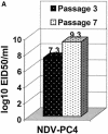
(A) Infectious titers of recombinant NDV-PC4 at passages 3 and 7. Titers were determined as described under the legend for Fig. 2. (B) Pathogenicity of NDV-PC4 at passage 3 (PC4-3) and passage 7 (PC4-7) in SPF chicken embryos. Mortality was determined as described in the legend for Fig. 3. (C) Editing frequency of NDV-PC4 at passages 3 (PC4-3) and passage 7 (PC4-7). mRNA editing was determined from independent clones (Table 1) as described under the legend for Fig. 4.
FIG. 5.
FIG. 5.
(A) Infectious titers of recombinant NDV-PC4 at passages 3 and 7. Titers were determined as described under the legend for Fig. 2. (B) Pathogenicity of NDV-PC4 at passage 3 (PC4-3) and passage 7 (PC4-7) in SPF chicken embryos. Mortality was determined as described in the legend for Fig. 3. (C) Editing frequency of NDV-PC4 at passages 3 (PC4-3) and passage 7 (PC4-7). mRNA editing was determined from independent clones (Table 1) as described under the legend for Fig. 4.
FIG. 6.
FIG. 6.
Comparison of putative mRNA-template hybrids of rNDV and NDV-T2 during the editing process. The template negative-strand genomes (top strands) were written 3′ to 5′, and the mRNA chains (bottom strands) were written 5′to 3′. The editing frequency of both viruses for P, V, and W mRNAs are shown in percentage. The only difference between the two viruses at position 2278 (C-U2278) was shown in boldface lowercase. The unique putative base pairings are underlined. As much as 85% of P-gene derived mRNAs in NDV-T2 are edited, whereas only 32% of the mRNAs in the parent virus represent V and W mRNAs.

Similar articles

Cited by

References

    1. Baron, M. D., and T. Barrett. 2000. Rinderpest viruses lacking the C and V proteins show specific defects in growth and transcription of viral RNAs. J. Virol. 74:2603-2611. - PMC - PubMed
    1. Beard, C. W., and R. P. Hanson. 1984. Newcastle disease, p. 452-470. In M. S. Hofstad, H. J. Barnes, B. W. Calnek, W. M. Reid, and H. W. Yoder (ed.), Diseases of poultry. Iowa State University Press, Ames.
    1. Buchholz, U. J., S. Finke, and K.-K. Conzelmann. 1999. Generation of bovine respiratory syncytial virus (BRSV) from cDNA: BRSV NS2 is not essential for virus replication in tissue culture, and the human RSV leader region acts as a functional BRSV genome promoter. J. Virol. 73:251-259. - PMC - PubMed
    1. Delenda, C., S. Hausmann, D. Garcin, and D. Kolakofsky. 1997. Normal cellular replication of Sendai virus without the trans-frame, nonstructural V protein. Virology 228:55-62. - PubMed
    1. Delenda, C., G. Taylor, S. Hausmann, D. Garcin, and D. Kolakofsky. 1998. Sendai viruses with altered P, V, and W protein expression. Virology 242:327-337. - PubMed

LinkOut - more resources