. 2003 Sep;77(17):9287-94.
doi: 10.1128/jvi.77.17.9287-9294.2003.

Human cytomegalovirus US2 causes similar effects on both major histocompatibility complex class I and II proteins in epithelial and glial cells

Affiliations

Human cytomegalovirus US2 causes similar effects on both major histocompatibility complex class I and II proteins in epithelial and glial cells

Nagendra R Hegde et al. J Virol. 2003 Sep.

Abstract

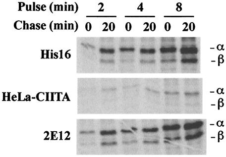
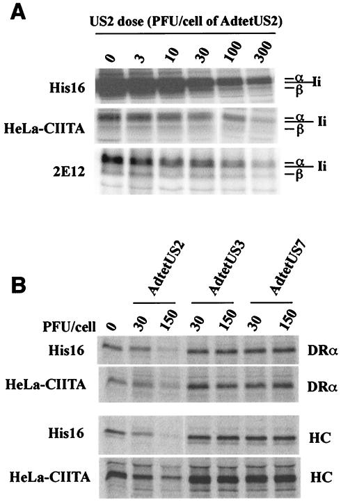
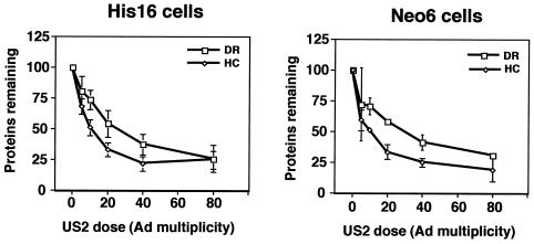
The human cytomegalovirus (HCMV) glycoprotein US2 specifically binds to major histocompatibility complex (MHC) class I heavy chain (HC) and class II proteins DRalpha and DMalpha, triggering their degradation by proteasomes. Effects of US2 on class II proteins were originally characterized in HCMV- or adenovirus vector-infected U373 astroglioma cells. Here, we have extended characterization of US2-mediated degradation of class II DRalpha to two other cell lines, including biologically relevant epithelial cells. Comparison of the effects of US2 in cells expressing both class I and II proteins demonstrated only a slight preference for class I HC. Moreover, US2 caused degradation of DRalpha and DMalpha when these proteins were expressed by transfection without DRbeta, invariant chain (Ii), or DMbeta. Therefore, US2 binds to alpha chains of DR and DM and triggers endoplasmic reticulum degradation without formation of class II DR alphabeta/Ii or DM alphabeta complexes. Similar levels of degradation of class II alpha were observed in cells expressing vastly different amounts of class II, suggesting that cellular factors, other than class II, were limiting. We concluded that US2 has broad effects in a variety of cells that express both class I and II proteins and is relevant to HCMV infection in vivo.


Figures

FIG. 1.
FIG. 1.
Expression of class II proteins by various cell lines. His16, HeLa-CIITA, and 2E12 cells were labeled in suspension for 2, 4, or 8 min with [35S]methionine-cysteine, and the label was chased for 0 or 20 min. NP-40-DOC cell extracts derived from 6 × 10⁶ (His16, 2E12) or 15 × 106 (HeLa-CIITA) cells were mixed with anti-DRα MAb DA6.147, and MHC class II complexes immunoprecipitated.
FIG. 2.
FIG. 2.
US2 causes loss of class I and II proteins. (A) His16, HeLa-CIITA, or 2E12 cells were left uninfected (0 PFU/cell) or were infected with AdtetUS2 and Adtet-trans using 3 and 0.6, 10 and 2, 30 and 6, 100 and 20, or 300 and 60 PFU/cell, respectively, for 18 h. Infected cells were labeled with [35S]methionine-cysteine for 3 h, and cell extracts were made using NP-40-DOC lysis buffer. Class II complexes were immunoprecipitated from 6 × 10⁶ (His16, 2E12) or 15 × 106 (HeLa-CIITA) cells using MAb DA6.147. (B) His16 (6 × 10⁶) and HeLa-CIITA (15 × 106) cells were left uninfected (0 PFU/cell) or were infected with AdtetUS2, AdtetUS3, or AdtetUS7, in each case with Adtet-trans using 30 and 6 PFU/cell or 150 and 30 PFU/cell, respectively, for 18 h. Infected cells were labeled with [35S]methionine-cysteine for 1 min, and the label was chased for 20 min. Cell extracts were made with 1% SDS lysis buffer and denatured at 100°C, and the SDS was diluted 10-fold with 1% Triton X-100 buffer. Lysates were divided equally into two, and class I HC or class II α chain (DRα) was immunoprecipitated with the MAb's HC10 or DA6.147, respectively.
FIG. 3.
FIG. 3.
US2 causes degradation of class II α when expressed without the β chain. HeLa cells were transiently transfected with plasmids containing either the class II α (HeLa/DRα) or β genes (HeLa/DRβ), or both plasmids (HeLa-DRα/DRβ), or one containing the DMα gene (HeLa-DMα). After 48 h, these transfected HeLa cells (4 × 10⁶) and His16 cells (1 × 10⁶) were left uninfected (0 PFU/cell) or infected with 3 and 0.6, 10 and 2, 30 and 6, 100 and 20, or 300 and 60 PFU/cell of AdtetUS2 and Adtet-trans, respectively, for 18 h. The cells were then labeled with [35S]methionine-cysteine for 3 h. Class II α was immunoprecipitated using MAb DA6.147, and class II β was immunoprecipitated with MAb HB10A. αβ complexes were precipitated from HeLa cells transfected with both DRα and DRβ plasmids with MAb DA6.147, and DMα was precipitated with MAb 5C1. Note that fewer His16 cells were used, and extracts of these cells were made using SDS buffer, as in Fig. 2, allowing selective precipitation of DRα but reducing binding of MAb DA6.147 compared with extracts of transfected HeLa cells made using NP-40-DOC buffer (see Fig. 4A).
FIG. 4.
FIG. 4.
Similar degradation in cells with different amounts of class II proteins. (A) His16 cells (1 × 10⁶) or DRα-transfected HeLa cells (4 × 10⁶) were labeled with [35S]methionine-cysteine for 3 h. His 16 cells were lysed in NP-40-DOC buffer (left lane denoted α/β/Ii)) or in buffer containing 1% SDS, and the extract was denatured at 100°C before the SDS was diluted with 1% Triton X-100 buffer (middle lane, denoted α). Transfected HeLa cells (HeLa-DRα) were lysed in NP-40-DOC buffer. All samples were immunoprecipitated with anti-DRα MAb DA6.147. (B) His16 or 2E12 cells (1 × 10⁶) or transfected HeLa cells (4 × 10⁶) were left uninfected (0 PFU/cell) or infected with 3 and 0.6, 10 and 2, 30 and 6, 100 and 20, or 300 and 60 PFU of AdtetUS2 and Adtet-trans/cell, respectively, for 18 h. Infected cells were labeled with [35S]methionine-cysteine for 3 h, extracts were made in every case with 1% SDS lysis buffer, proteins were denatured, and the SDS was diluted with 1% Triton X-100 buffer. Class II α or β chains were immunoprecipitated from the cell extracts with anti-α MAb DA6.147 or anti-β MAb HB10A, respectively. The intensities of protein bands were quantified by phosphorimager analyses. The data are the averages of three independent experiments, and the density of protein bands obtained from cells left uninfected (no AdtetUS2) was considered 100%. Data points without error bars are due to standard deviations that are too small for depiction. (C) Cells were labeled as in panel B, and US2 was immunoprecipitated with rabbit polyclonal serum to an N-terminal peptide of US2. The amount of US2 was quantified by phosphorimager analyses from the three experiments depicted in panel A. (D) The effects of US2 on class II DMα were analyzed as for panel B, except that HeLa cells were transfected with a plasmid containing the DMα gene and compared to His16 cells that also express DM. Immunoprecipitation was performed with anti-DMα MAb 5C1.
FIG. 5.
FIG. 5.
US2 shows preference for class I HC over class II α chain. His16 or Neo6 cells (6 × 10⁶) were left uninfected (0 PFU/cell) or infected with 5 and 1, 10 and 2, 20 and 4, 40 and 8, or 80 and 16 PFU/cell of AdtetUS2 and Adtet-trans, respectively, for 18 h. Cells in suspension were labeled for 1 min, the label was chased for 20 min, cell extracts were prepared using SDS lysis buffer, proteins were denatured, and the SDS was diluted as described in the legend for Fig. 4. Extracts were then divided equally into two, and class II α (DRα) or class I HC was immunoprecipitated with MAbs DA6.147 or HC10, respectively. The amount of α or β that remained after expression of each dose of US2 expression was quantified by phosphorimager analyses and compared to that in uninfected cells (100%). The data represent the averages of three independent experiments. Data points without error bars are due to standard deviations that are too small for depiction.
FIG. 6.
FIG. 6.
Different Ad vectors expressing US2 cause degradation of MHC proteins. His16 cells (6 × 10⁶) were infected with 50 PFU/cell of Ad viruses AdUS2 and AdUS11 (described by Rehm et al. [43]) or Adβ-gal (described by Tomanin et al. [49]), or AdtetUS2, AdtetUS3, or AdtetUS11 (described elsewhere by our group [23, 50]). The cells were labeled in suspension with [35S]methionine-cysteine for 1 min, and the label was chased for 20 min. Cell extracts were made using SDS lysis buffer, proteins were denatured, and the SDS was diluted as described in the legend for Fig. 4. Extracts were divided into three, and class II α chain (DRα), class I HC (panel A), or transferrin receptor (TfR; panel B) were immunoprecipitated with MAbs DA6.147, HC10, or B3/25, respectively.

References

    1. Ahn, K., A. Angulo, P. Ghazal, P. A. Peterson, Y. Yang, and K. Fruh. 1996. Human cytomegalovirus inhibits antigen presentation by a sequential multistep process. Proc. Natl. Acad. Sci. USA 93:10990-10995. - PMC - PubMed
    1. Ahn, K., A. Gruhler, B. Galocha, T. R. Jones, E. J. Wiertz, H. L. Ploegh, P. A. Peterson, Y. Yang, and K. Fruh. 1997. The ER-luminal domain of the HCMV glycoprotein US6 inhibits peptide translocation by TAP. Immunity 6:613-621. - PubMed
    1. Ben-Arieh, S. V., B. Zimerman, N. I. Smorodinsky, M. Yaacubovicz, C. Schechter, I. Bacik, J. Gibbs, J. R. Bennink, J. W. Yewdell, J. E. Coligan, H. Firat, F. Lemonnier, and R. Ehrlich. 2001. Human cytomegalovirus protein US2 interferes with the expression of human HFE, a nonclassical class I major histocompatibility complex molecule that regulates iron homeostasis. J. Virol. 75:10557-10562. - PMC - PubMed
    1. Bitmansour, A. D., S. L. Waldrop, C. J. Pitcher, E. Khatamzas, F. Kern, V. C. Maino, and L. J. Picker. 2001. Clonotypic structure of the human CD4+ memory T cell response to cytomegalovirus. J. Immunol. 167:1151-1163. - PubMed
    1. Borza, C. M., and L. M. Hutt-Fletcher. 2002. Alternate replication in B cells and epithelial cells switches tropism of Epstein-Barr virus. Nat. Med. 8:594-599. - PubMed

Publication types

MeSH terms