. 2003 Sep;77(17):9295-304.
doi: 10.1128/jvi.77.17.9295-9304.2003.

Heterologous human immunodeficiency virus type 1 lentiviral vectors packaging a simian immunodeficiency virus-derived genome display a specific postentry transduction defect in dendritic cells

Affiliations

Heterologous human immunodeficiency virus type 1 lentiviral vectors packaging a simian immunodeficiency virus-derived genome display a specific postentry transduction defect in dendritic cells

Caroline Goujon et al. J Virol. 2003 Sep.

Abstract

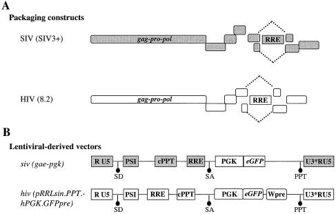
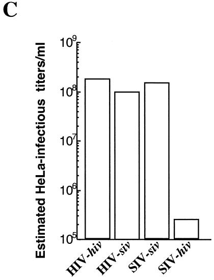
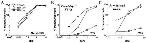
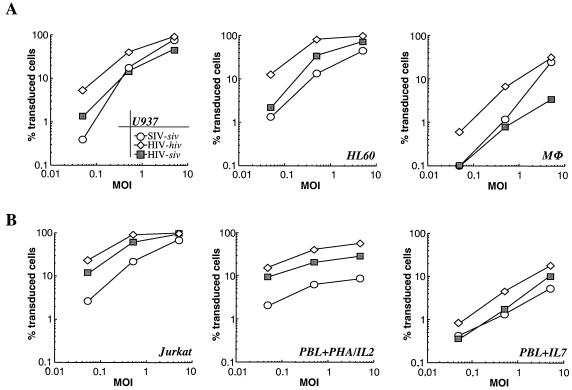
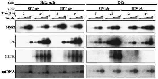
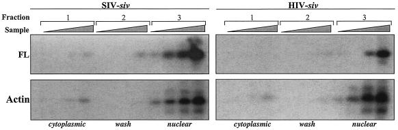
Heterologous lentiviral vectors (LVs) represent a way to address safety concerns in the field of gene therapy by decreasing the possibility of genetic recombination between vector and packaging constructs and the generation of replication-competent viruses. Using described LVs based on human immunodeficiency virus type 1 (HIV-1) and simian immunodeficiency virus MAC251 (SIV(MAC251)), we asked whether heterologous virion particles in which trans-acting factors belonged to HIV-1 and cis elements belonged to SIV(MAC251) (HIV-siv) would behave as parental homologous vectors in all cell types. To our surprise, we found that although the heterologous HIV-siv vector was as infectious as its homologous counterpart in most human cells, it was defective in the transduction of dendritic cells (DCs) and, to a lesser extent, macrophages. In DCs, the main postentry defect was observed in the formation of two-long-terminal-repeat circles, despite the fact that full-length proviral DNA was being synthesized and was associated with the nucleus. Taken together, our data suggest that heterologous HIV-siv vectors display a cell-dependent infectivity defect, most probably at a post-nuclear entry migration step. As homologous HIV and SIV vectors do transduce DCs, we believe that these results underscore the importance of a conserved interaction between cis elements and trans-acting viral factors that is lost or suboptimal in heterologous vectors and essential only in the transduction of certain cell types. For gene therapy purposes, these findings indicate that the cellular tropism of LVs can be modulated not only through the use of distinct envelope proteins or tissue-specific promoters but also through the specific combinatorial use of packaging and transfer vector constructs.


Figures

FIG. 1.
FIG. 1.
Schematic representation of the SIVMAC251- and HIV-1-based LVs used. Structures of the SIVMAC251 and HIV-1 packaging constructs (A) and of the respective vector genomes (B) carrying an eGFP reporter gene under the control of the phosphoglycerate kinase 1 promoter (PGK). Original names of the constructs are reported within parentheses (capital letters for the packaging constructs, italic small letters for the vector genomes). SD and SA, major splice donor and acceptor sites, respectively. The U3s of both siv and hiv are self-inactivating and result in an impaired U3 viral promoter (U3*). cPPT actually denotes a composite cPPT-CTS element (10, 21, 54). Accessory genes carried by the packaging constructs are not detailed here. (C) Viral preparations were normalized by exogenase reverse transcriptase activity and used to transduce HeLa cells. The estimated infectious titers per milliliter are reported.
FIG. 1.
FIG. 1.
Schematic representation of the SIVMAC251- and HIV-1-based LVs used. Structures of the SIVMAC251 and HIV-1 packaging constructs (A) and of the respective vector genomes (B) carrying an eGFP reporter gene under the control of the phosphoglycerate kinase 1 promoter (PGK). Original names of the constructs are reported within parentheses (capital letters for the packaging constructs, italic small letters for the vector genomes). SD and SA, major splice donor and acceptor sites, respectively. The U3s of both siv and hiv are self-inactivating and result in an impaired U3 viral promoter (U3*). cPPT actually denotes a composite cPPT-CTS element (10, 21, 54). Accessory genes carried by the packaging constructs are not detailed here. (C) Viral preparations were normalized by exogenase reverse transcriptase activity and used to transduce HeLa cells. The estimated infectious titers per milliliter are reported.
FIG. 2.
FIG. 2.
Cell-specific infectivity defect of HIV-siv vectors in DCs. VSVg (A and B)- and JR-FL (C)-pseudotyped LVs were produced by calcium phosphate DNA transfection into 293Tcells and purified by ultracentrifugation through sucrose, and their infectious titers were determined upon transduction of target HeLa or 293T cells. HeLa cells (105) and DCs were then transduced in parallel with logarithmic dilutions of homologous SIV-siv and HIV-hiv vectors along with the heterologous HIV-siv vector. Successful transduction events were analyzed by flow cytometry by determining the percentage of eGFP-positive cells 72 to 96 h posttransduction. Results obtained from a representative experiment are shown here, with the percentages of transduced eGFP-positive cells as the ordinates and the MOIs as the abscissas.
FIG. 3.
FIG. 3.
Transduction of primary cells and different cell lines with homologous HIV, SIV, and heterologous HIV-siv LVs. Virions, produced as described in the legend to Fig. 2, were used to transduce monocyte/macrophage-like cell lines and primary macrophages (A) and lymphoid Jurkat cells along with primary PBLs stimulated with PHA-IL-2 or IL-7 (B). Results from representative experiments are depicted. The percentages of transduced eGFP-positive cells (ordinates) are shown as a function of MOIs (abscissas).
FIG. 4.
FIG. 4.
Characterization of HIV-siv virion particles. (A) Western blot analysis of cell lysates (cell-assoc.; lanes 1 to 3) and virion particles (virion-assoc.; lanes 4 to 6) produced after calcium phosphate transfection of DNA into 293T cells. Virion particles were purified onto a double-step sucrose gradient prior to analysis. Western blots were probed by using an anti-CA antibody that recognizes a common epitope present on both HIV and SIV CA and with an antibody that recognizes the VSVg envelope, as indicated. The positions of migration of molecular mass markers (in kilodaltons) are indicated on the right. The products generated upon viral protease processing of SIV (p57-p45-p27) and HIV-1 (p55-p41-p24) Gag polyproteins and recognized by the anti-CA antibody are shown on the left. (B) Genomic-vector RNA incorporation as determined by slot blot analysis on normalized amounts of virion particles by using a probe that hybridizes to the common eGFP gene sequence present on both siv and hiv vector genomes. RNase A treatment of viral RNA preparations was included as a control for DNA contaminations. Twofold dilutions of viral RNA were used as a control for the linearity of the assay. Std. dilutions, standard dilutions.
FIG. 5.
FIG. 5.
Semiquantitative PCR analysis of reverse transcription intermediates upon cell transduction. Normalized amounts of SIV-siv and HIV-siv vectors were used to transduce 105 HeLa cells and DCs, as indicated. Cell aliquots were harvested at 2 and 24 h posttransduction, lysed, and analyzed by PCR. PCR analysis was conducted with fivefold serial dilutions of starting material by using primers that recognized specifically the MSSS, the FL, and the 2LTR forms produced during the reverse transcription process, as indicated. Amplification of mitochondrial DNA (mtDNA) was used for DNA input and quality control. PCR products were transferred onto a nylon membrane and hybridized with P32-labeled specific internal probes prior to phosphorimager analysis.
FIG. 6.
FIG. 6.
Subcellular fractionation and localization of proviral DNA in DCs. DCs were transduced with SIV-siv and HIV-siv vectors and fractioned 24 h later into cytoplasmic (fraction 1), wash (fraction 2), and nuclear (fraction 3) fractions, as indicated. PCR was then conducted with serial dilutions of samples by using a primer specific for FL proviral DNA and one specific for actin as a control for nuclear integrity. The amplification products obtained after PCR analysis were transferred onto a nylon membrane and hybridized with P32-labeled specific internal probes prior to phosphorimager analysis.

Similar articles

Cited by

References

    1. Basu, S., and H. E. Varmus. 1990. Moloney murine leukemia virus integration protein produced in yeast binds specifically to viral att sites. J. Virol. 64:5617-5625. - PMC - PubMed
    1. Berthoux, L., G. J. Towers, C. Gurer, P. Salomoni, P. P. Pandolfi, and J. Luban. 2003. As(2)O(3) enhances retroviral reverse transcription and counteracts Ref1 antiviral activity. J. Virol. 77:3167-3180. - PMC - PubMed
    1. Besnier, C., Y. Takeuchi, and G. Towers. 2002. Restriction of lentivirus in monkeys. Proc. Natl. Acad. Sci. USA 99:11920-11925. - PMC - PubMed
    1. Best, S., P. Le Tissier, G. Towers, and J. P. Stoye. 1996. Positional cloning of the mouse retrovirus restriction gene Fv1. Nature 382:826-829. - PubMed
    1. Braaten, D., E. K. Franke, and J. Luban. 1996. Cyclophilin A is required for an early step in the life cycle of human immunodeficiency virus type 1 before the initiation of reverse transcription. J. Virol. 70:3551-3560. - PMC - PubMed

Publication types