. 2003 Sep;77(17):9346-58.
doi: 10.1128/jvi.77.17.9346-9358.2003.

Activation of mitogen-activated protein kinase and NF-kappaB pathways by a Kaposi's sarcoma-associated herpesvirus K15 membrane protein

Affiliations

Activation of mitogen-activated protein kinase and NF-kappaB pathways by a Kaposi's sarcoma-associated herpesvirus K15 membrane protein

Melanie M Brinkmann et al. J Virol. 2003 Sep.

Abstract

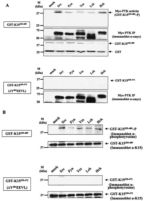
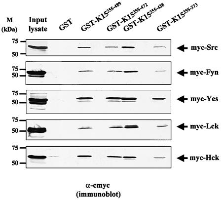
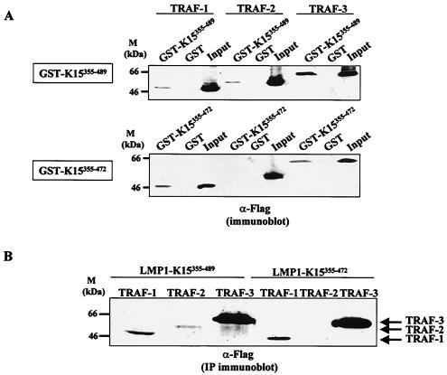
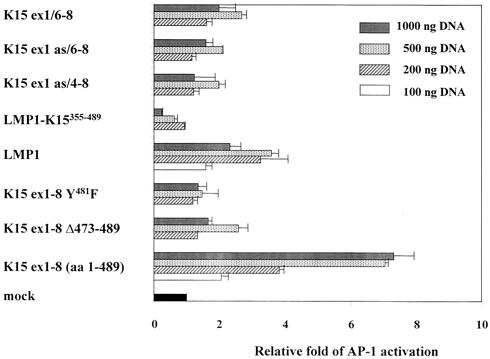
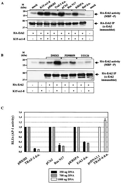
The K15 gene of Kaposi's sarcoma-associated herpesvirus (also known as human herpesvirus 8) consists of eight alternatively spliced exons and has been predicted to encode membrane proteins with a variable number of transmembrane regions and a common C-terminal cytoplasmic domain with putative binding sites for SH2 and SH3 domains, as well as for tumor necrosis factor receptor-associated factors. These features are reminiscent of the latent membrane proteins LMP-1 and LMP2A of Epstein-Barr virus and, more distantly, of the STP, Tip, and Tio proteins of the related gamma(2)-herpesviruses herpesvirus saimiri and herpesvirus ateles. These viral membrane proteins can activate a number of intracellular signaling pathways. We have therefore examined the abilities of different K15-encoded proteins to initiate intracellular signaling. We found that a 45-kDa K15 protein derived from all eight K15 exons and containing 12 predicted transmembrane domains in addition to the cytoplasmic domain activated the Ras/mitogen-activated protein kinase (MAPK) and NF-kappaB pathways, as well as (more weakly) the c-Jun N-terminal kinase/SAPK pathway. Activation of the MAPK and NF-kappaB pathways required phosphorylation of tyrosine residue 481 within a putative SH2-binding site (YEEVL). This motif was phosphorylated by the tyrosine kinases Src, Lck, Yes, Hck, and Fyn. The region containing the YEEVL motif interacted with tumor necrosis factor receptor-associated factor 2 (TRAF-2), and a dominant negative TRAF-2 mutant inhibited the K15-mediated activation of the Ras/MAPK pathway, suggesting the involvement of TRAF-2 in the initiation of these signaling routes. In contrast, several smaller K15 protein isoforms activated these pathways only weakly. All of the K15 isoforms tested were, however, localized in lipid rafts, suggesting that incorporation into lipid rafts is not sufficient to initiate signaling. Additional regions of K15, located presumably in exons 2 to 5, may therefore contribute to the activation of these pathways. These findings illustrate that the 45-kDa K15 protein engages pathways similar to LMP1, LMP2A, STP, Tip, and Tio but combines functional features that are separated between LMP1 and LMP2A or STP and Tip.


Figures

FIG. 1.
FIG. 1.
ORF K15 expression constructs used in this study and their putative protein products. The K15 ORF is multiply and alternatively spliced (as). The major transcript identified in PEL cells by RT-PCR is fully spliced and contains all eight exons (K15 ex1-8; aa 1 to 489). It encodes a membrane protein with up to 12 transmembrane domains and a cytoplasmic C-terminal domain (aa 355 to 489). The C-terminal domain contains motifs reminiscent of SH2, SH3, and TRAF-like binding sites. The distal TRAF-like binding site and the distal SH2-binding motif Y481EEVL are deleted in construct K15 ex1-8 Δ473 to 489. K15 ex1-8 Y481F carries a point mutation in the distal SH2-binding motif (Y481→F481EEVL). The LMP1-K15355-489 chimera was constructed by fusing the six transmembrane domains of LMP1 to the cytoplasmic C-terminal end (aa 355 to 489) of K15. The splice variants K15 ex1/6-8, K15 ex1 as/6-8, and K15 ex1 as/4-8 differ in the number of transmembrane domains they contain, but all contain the C-terminal domain.
FIG. 2.
FIG. 2.
The 45-kDa K15 protein (aa 1 to 489) is associated with lipid rafts. Cos7 cells were transiently transfected with K15 expression construct K15 ex1-8 (A); the natural splice variant K15 ex1/6-8 (B), K15 ex1 as/4-8 (C), or K15 ex1 as/6-8 (D); or the LMP1-K15355-489 chimera (E). At 48 h after transfection, Cos7 cells were lysed in TNE buffer and extracts were analyzed on a flotation sucrose gradient as described in Materials and Methods. After ultracentrifugation, 1-ml fractions were collected starting at the top of the gradient and analyzed by Western blotting with a rabbit antibody to K15. Lipid raft-associated proteins are localized at the interface of 5 and 35% sucrose (fractions 4 and 5). Soluble proteins and solubilized membrane proteins of the nonraft plasma membrane are localized to the higher-density sucrose fractions (fractions 10 to 12, 35 to 42.5% sucrose). P, pellet. (A, top) The 45-kDa form of K15 ex1-8 (aa 1 to 489) localizes to lipid rafts (fractions 4 and 5, interface of 5 and 35% sucrose) and to the fractions containing solubilized membrane proteins (fractions 10 to 12, 35 to 42.5% sucrose). (A, bottom) Western blotting of the samples from the upper part of this panel probed with anti-caveolin 1. Endogenous caveolin 1 served as a positive control for lipid raft localization. (B) The 33- to 35-kDa protein derived from splice variant K15 ex1/6-8 is localized in the lipid raft fraction and high-density sucrose fractions (fractions 8 to 11). *, possible dimeric form. (C) The ∼21-kDa protein derived from splice variant K15 ex1 as/4-8 (see text) is found in lipid rafts and high-density sucrose fractions. (D) The ∼21-kDa protein derived from K15 ex1 as/6-8 is localized in lipid rafts and also in fractions containing solubilized membrane proteins (see text). (E) The LMP1-K15355-489 chimera is found in lipid rafts and high-density sucrose fractions.
FIG. 3.
FIG. 3.
The tyrosine residue of the distal SH2-binding motif (Y481EEVL) of the K15 C-terminal domain is phosphorylated by members of the Src kinase family of PTKs. Purified GST-K15355-489 and GST-K15355-472 proteins were used as substrates in in vitro kinase assays with immunoprecipitated (IP) myc-tagged PTKs and [γ-32P]ATP. (A) The PTK family members Src, Fyn, Yes, Lck, and Hck phosphorylate the C-terminal domain of K15 in an in vitro immunocomplex kinase assay (top, upper part). When the last 17 aa encompassing the SH2-binding motif (Y481EEVL) of the C terminus were deleted (GST-K15355-472), no phosphorylation was observed (bottom, upper part). The expression level of the myc-tagged immunoprecipitated Src kinases was detected with an anti-c-myc antibody (top, middle part, and bottom, lower part). The amounts of GST-K15355-489 fusion protein and nonfused GST protein used in the in vitro kinase reactions were also analyzed with a Coomassie-stained SDS gel (top, lower part). (B) The kinase reaction mixtures shown in panel A were analyzed by Western blotting with an anti-phosphotyrosine antibody. Expression of the GST-K15 fusion proteins was detected with an anti-K15 monoclonal antibody.
FIG. 4.
FIG. 4.
The nonreceptor PTKs Src, Fyn, Yes, Lck, and Hck bind to the C-terminal domain of K15. In a GST pulldown experiment, the GST-K15355-489, GST-K15355-472, GST-K15355-438, and GST-K15355-373 fusion proteins immobilized on glutathione Sepharose beads were incubated with lysates of HEK 293-T cells transfected with expression vectors for myc-tagged PTKs. Bound PTKs were detected by Western blotting with an antibody to c-myc.
FIG. 5.
FIG. 5.
Interaction of TRAF-1, -2, and -3 with the carboxy-terminal domain of K15. (A) Flag-tagged cDNA expression constructs of TRAF-1, -2, and -3 were transiently transfected into HEK 293 cells, and the interaction of the TRAF proteins with the carboxy-terminal domain of K15 (aa 355-489) was examined by GST pulldown assays as described in Materials and Methods. (Top) Interaction of TRAF with the entire carboxy-terminal domain of K15. Lanes: 1 to 3, TRAF-1-transfected cells; 4 to 6, TRAF-2-transfected cells; 7 to 9, TRAF-3-transfected cells; 1, 4, and 7, TRAF proteins bound to GST-K15355-489; 2, 5, and 8, proteins bound to GST; 3, 6, and 9, input cell lysates. (Bottom) Interaction with the carboxy-terminal region of K15 from which the terminal 17 aa, containing the distal TRAF-like binding site, have been deleted (GST-K15355-472). Samples were loaded as in the upper part of this panel. (B) HEK 293 cells were transiently cotransfected with Flag-tagged TRAF-1 (lanes 1 and 4), TRAF-2 (lanes 2 and 5), and TRAF-3 (lanes 3 and 6) expression constructs together with the LMP1-K15355-489 (lanes 1 to 3) or LMP1-K15355-472 (lanes 4 to 6) chimera. At 48 h after transfection, detergent-extracted cell lysates were immunoprecipitated (IP) with rabbit anti-K15 serum and analyzed by Western blotting, followed by staining with an anti-Flag monoclonal antibody.
FIG. 6.
FIG. 6.
The 45-kDa K15 protein activates the transcription factor NF-κB in a luciferase-based reporter assay. HEK 293-T cells were transiently cotransfected with 50 ng of the luciferase reporter plasmid p3EnhκBconA-Luc containing three NF-κB binding sites upstream of the luciferase gene and different amounts of K15 expression constructs (200 and 500 ng of DNA). At 24 h after transfection, cells were lysed and analyzed for luciferase activity. Shown is the relative fold activation compared to that of empty-vector (mock)-transfected (200 and 500 ng of DNA) cells based on triplicate samples. Equal expression levels of K15 proteins were analyzed by Western blotting with rabbit K15 antiserum (data not shown).
FIG. 7.
FIG. 7.
Transcription factor AP-1 is activated by K15. HEK 293-T cells were transiently cotransfected with the AP-1 luciferase reporter plasmid pRTU14 containing four AP-1-binding sites upstream of the luciferase gene and different amounts of K15 expression constructs (100, 200, 500, and 1,000 ng). After transfection, cells were grown in medium containing 1% FCS, lysed after 24 h, and analyzed for luciferase activity. Shown is the relative fold activation compared to that of mock-transfected (100, 200, 500, and 1,000 ng) cells based on triplicate samples. Equal expression levels of K15 proteins were analyzed by Western blotting with rabbit K15 antiserum (data not shown).
FIG. 8.
FIG. 8.
Immunocomplex kinase assays with MAPKs Erk2 and JNK1. HEK 293-T cells were transiently cotransfected with 1 μg of the HA-tagged MAPK Erk2 or JNK1 and 1 μg of the K15 (Fig. 1) or LMP1 expression construct. After transfection, cells were grown in medium containing 1% FCS and lysed after 24 h in TBS-T buffer containing phosphatase inhibitors. The cell lysates were then subjected to immunoprecipitation with HA-antibody 12C5 coupled to protein G beads overnight. MBP or purified GST-c-Jun fusion protein served as the substrate in the in vitro kinase reaction mixture with Erk2 (A) or JNK1 (B), respectively. The immunoprecipitated (IP) kinase was incubated with its substrate and [γ-32P]ATP for 30 min at 25°C in kinase reaction buffer and subsequently analyzed by autoradiography (top of panels A and B). Western blots of the kinase reactions were probed with specific Erk2 or JNK1 antibodies to ensure equal expression levels of the MAPKs (middle of panels A and B). Cell lysates were analyzed for equal expression of K15 constructs by Western blotting and probing with a K15 antibody (bottom of panels A and B).
FIG.9.
FIG.9.
Dominant negative-negative (d.n.) mutant forms of TRAF-2, Erk2, Raf, and Ras and the MEK1/2 inhibitors UO126 and PD98059 reduce the Erk2 and AP1 activation induced by K15. (A) Seven hundred nanograms of a dominant negative mutant form of TRAF-2, Ras, Raf, or Erk2 or the respective empty expression vector, pRKH5, pCis2, or pKRSPA, was cotransfected with 1 μg of HA-Erk2 and 1 μg of K15 ex1-8 (aa 1 to 489) or empty K15 expression vector (mock) where indicated. Erk2 kinase activity was monitored by phosphorylation of MBP (top). The Western blot probed with anti-Erk2 antibody shows equal expression levels of the Erk2 kinase for all samples. (B) HEK 293-T cells were cotransfected with 1 μg of HA-Erk2 and 1 μg of K15 ex1-8 (aa 1 to 489) expression construct or empty K15 expression vector. At 14 h after transfection, DMSO alone or the MEK1/2 inhibitor PD98059 or UO126 was added to the medium at 50 μM from a 50 mM stock solution in DMSO. Cells were incubated for a further 8 h before protein extraction. In vitro kinase assays were performed as described in the legend to Fig. 8 and Materials and Methods. The Western blot probed with anti-Erk2 antibody shows equal expression levels of the Erk2 kinase for all samples. (C) AP-1 luciferase-based reporter assay. HEK 293-T cells were transiently cotransfected with 50 ng of the AP-1 reporter plasmid (pRTU14), 500 ng of the K15 ex1-8 (aa 1 to 489) expression construct, and 350, 700, or 1,000 ng of a d.n. mutant form of Ras, TRAF-2, TRAF-6, or Erk2 or the respective empty expression vector, pCis2, pRKH5, pcDNA3.1, or pKRSPA. Shown are the luciferase values (in relative light units) of the different dominant negative mutant forms with respect to those of the corresponding empty expression vectors, which were set at 1.

References

    1. Albrecht, J.-C., U. Friedrich, C. Kardinal, J. Koehn, B. Fleckenstein, S. M. Feller, and B. Biesinger. 1999. Herpesvirus ateles gene product Tio interacts with non-receptor protein tyrosine kinases. J. Virol. 73:4631-4639. - PMC - PubMed
    1. Alessi, D. R., A. Cuenda, P. Cohen, D. T. Dudley, and A. R. Saltiel. 1995. PD 098059 is a specific inhibitor of the activation of mitogen-activated protein kinase kinase in vitro and in vivo. J. Biol. Chem. 270:27489-27494. - PubMed
    1. Baud, V., Z. G. Liu, B. Bennett, N. Suzuki, Y. Xia, and M. Karin. 1999. Signaling by proinflammatory cytokines: oligomerization of TRAF2 and TRAF6 is sufficient for JNK and IKK activation and target gene induction via an amino-terminal effector domain. Genes Dev. 13:1297-1308. - PMC - PubMed
    1. Biesinger, B., A. Y. Tsyganov, H. Fickenscher, F. Emmrich, B. Fleckenstein, J. B. Bolen, and B. M. Bröker. 1995. The product of the herpesvirus saimiri open reading frame 1 (Tip) interacts with T-cell specific kinase p56lck in transformed cells. J. Biol. Chem. 270:4729-4734. - PubMed
    1. Blasig, C., C. Zietz, B. Haar, F. Neipel, S. Esser, N. H. Brockmeyer, E. Tschachler, S. Colombini, B. Ensoli, and M. Sturzl. 1997. Monocytes in Kaposi's sarcoma lesions are productively infected by human herpesvirus 8. J. Virol. 71:7963-7968. - PMC - PubMed

Publication types

MeSH terms