. 2003 Sep;77(17):9369-77.
doi: 10.1128/jvi.77.17.9369-9377.2003.

Passive immunization with neutralizing antibodies interrupts the mouse mammary tumor virus life cycle

Affiliations

Passive immunization with neutralizing antibodies interrupts the mouse mammary tumor virus life cycle

M Mpandi et al. J Virol. 2003 Sep.

Abstract

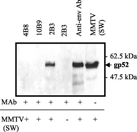
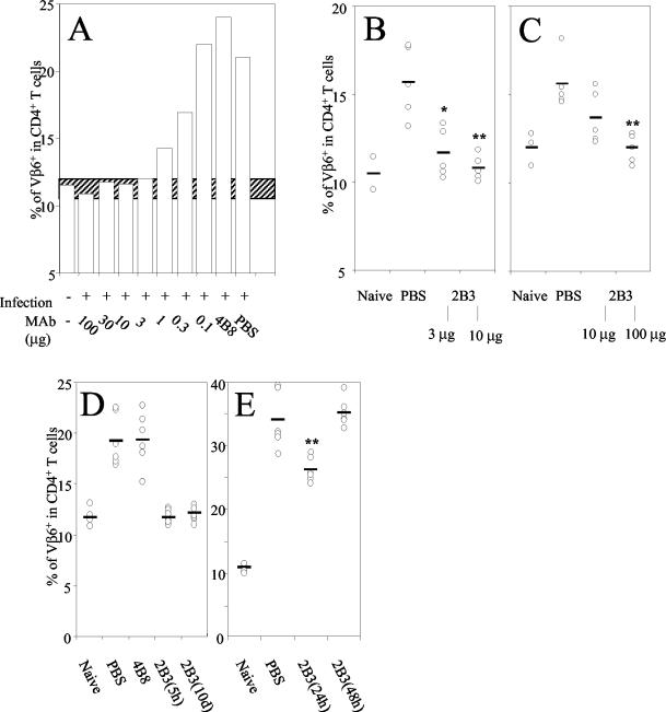
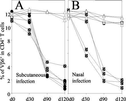
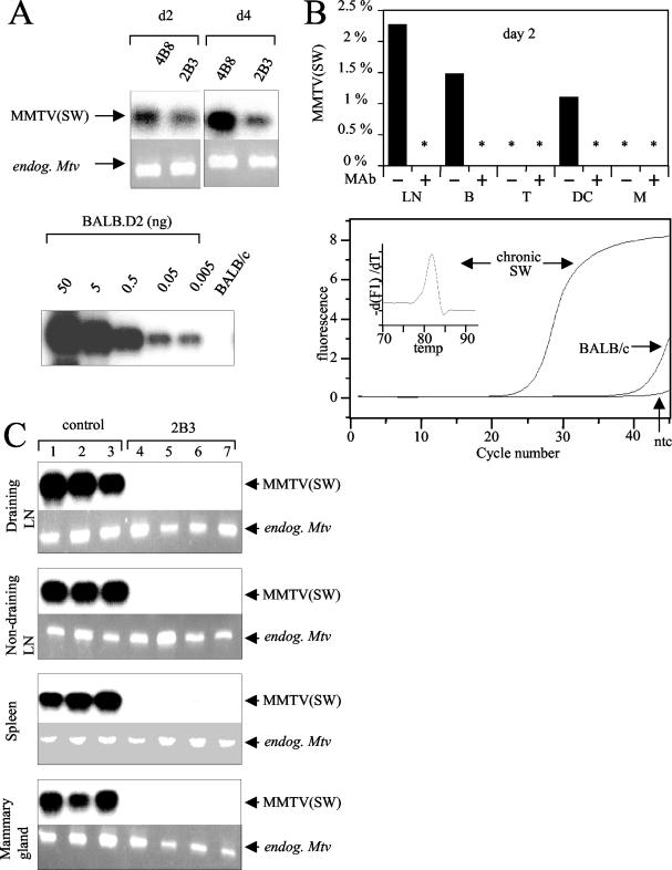
Mouse mammary tumor virus (MMTV) infects the host via mucosal surfaces and exploits the host immune system for systemic spread and chronic infection. We have tested a neutralizing rat monoclonal antibody specific for the retroviral envelope glycoprotein gp52 for its efficiency in preventing acute and chronic mucosal and systemic infection. The antibody completely inhibits the superantigen response and chronic viral infection following systemic or nasal infection. Surprisingly however, the antibody only partially inhibits the early infection of antigen-presenting cells in the draining lymph node. Despite this initially inefficient protection from infection, superantigen-specific B- and T-cell responses and systemic viral spread are abolished, leading to complete clearance of the retroviral infection and hence interruption of the viral life cycle. In conclusion, systemic neutralizing monoclonal antibodies can provide an efficient protection against chronic retroviral amplification and persistence.


Figures

FIG. 1.
FIG. 1.
Immunoprecipitation of MMTV SW with 4B8 (IgG2a[κ]) (lane 1 [counting from the left]), 10B9 (IgG2a[λ]) (lane 2), and 2B3 (IgG2a[κ]) (lane 3). Controls included Ab 2B3 without virus (lane 4), MMTV incubated with goat anti-gp52 (lane 5), and native MMTV (lane 6).
FIG. 2.
FIG. 2.
(A) Female adult BALB/c mice were i.p injected with different doses (100 to 0.1 μg) of MAb 2B3, the isotype-matched 4B8 MAb, or PBS and challenged by s.c. injection of MMTV SW 5 h later. Four days after infection lymphocytes isolated from draining LNs were analyzed by flow cytometry using anti-Vβ6-FITC and anti-CD4-PE MAbs. Data are mean values of Vβ6 expression on gated viable CD4+ T cells from two to four LNs. This assay was done twice with similar results. The hatched bar represents Vβ6 percentages in control BALB/c mice ±2 standard deviations. (B) Adult female mice were i.p. immunized with 3 or 10 μg of MAb 2B3. Control mice were injected with PBS. Twelve hours later mice were challenged nasally with MMTV SW. Six days later the percentage of Vβ CD4+ T cells in NALT was determined by flow cytometry. *, P < 0.01; **, P < 0.005. (C) Seven-day-old mice were i.p. injected with 10 or 100 μg of MAb 2B3 or PBS. Twelve hours later mice were infected by foster nursing on MMTV SW-infected lactating females. Seven days later, the percentage of Vβ+ CD4+ cells in PP from these mice was determined by flow cytometry. **, P < 0.005. (D) Female BALB/c mice were i.p. injected with 10 μg of MAb 2B3, control MAb 4B8, or PBS. After 5 h or 10 days mice were challenged by s.c. injection with MMTV SW into the hind footpad. Four days later the draining popliteal LNs were isolated and cells were analyzed by flow cytometry. The mean values and values for each individual experiment measuring Vβ6+ cells among CD4+ T cells in single draining LNs are shown. Four to six animals were analyzed separately in each group. The assay was done two to four times with similar results. *, P < 0.001. (E) Mice were s.c. infected with MMTV SW and 24 or 48 h later 10 μg of MAb 2B3 was injected i.p. The mean values and values for each experiment measuring Vβ6+ CD4+ T cells from six individual draining LNs are shown.
FIG. 3.
FIG. 3.
Female adult mice were i.p. immunized with 100 μg of MAb 2B3. As controls, mice were injected with the same dose of isotype-matched MAb 4B8 or PBS. After 12 h mice were challenged s.c. with MMTV SW in the left hind footpad (A) or via the nasal route (B). At the indicated time points the percentage of CD4+ Vβ6+ T cells in PBLs was determined by flow cytometry. Results for individual mice are shown. Boxes, PBS; circles, 4B8; triangles, 2B3.
FIG. 4.
FIG. 4.
Female adult mice were i.p. immunized with 100 μg of MAb 2B3. As controls, mice were injected with the isotype-matched MAb 4B8 or PBS. After 12 h mice were s.c. challenged with MMTV SW in the hind footpad. Two or 4 days later the draining popliteal LNs were removed. (A, top) Integrated copies of MMTV SW viruses on days 2 and 4 were detected by PCR using primers specific for Mtv-7 and the exogenous MMTV SW SAg. Analysis with primers specific for endogenous Mtv-6, -8, and -9 sequences served as internal controls to confirm equal amounts of DNA in each sample. (Bottom) To semiquantify infection levels, PCR with the identical MMTV SW primers was performed with serial dilutions of BALB.D2 mouse DNA in BALB/c mouse DNA. (B, top) Levels of MMTV SW infection on day 2 were quantified by real-time PCR in total LN cells and sorted B cells (B), T cells (T), DC, and macrophages (M). Values for MMTV SW infection were normalized with respect to D12Mit199values and are presented as percentages of the chronic infection level. Duplicate analysis showed that measurement errors for MMTV SW and D12Mit199 are 35 and 3%, respectively. *, samples negative for the MMTV SW sequence. (Bottom) The specificity of MMTV SW amplification was evaluated by comparing amplification plots of nontemplate control (ntc) and genomic DNA extracted from a chronically MMTV SW-infected BALB/c mouse (chronic SW) and a noninfected BALB/c mouse. In addition specificity was shown by the melting curve. (C) Female adult mice were passively immunized (i.p.) with 100 μg of 2B3 followed by challenge with MMTV SW s.c. 2 (lanes 1 to 6) or 11 months (lane 7) later. Draining popliteal LNs, nondraining LNs, spleens, and mammary glands were removed. DNA was analyzed by PCR with primers specific for the exogenous MMTV SW SAg and Mtv-7 as well as primers specific for the endogenous Mtvs.
FIG. 5.
FIG. 5.
Flow cytometry analysis of lymphocytes isolated from the draining popliteal LN 6 days after MMTV SW infection. (A) Among naive mice, 5.4% of all MHC-II+ cells express low levels of B220 (B220low B cells). In MMTV SW-infected mice, the percentage of MHC-II+ B220low B cells increases to 17.3%. In 2B3-treated infected mice, B220 expression (6.4%) is comparable to that in uninfected control mice. (B) The percentages of CD138-expressing cells gated on MHC-II+ B220low B cells are shown. (C) The percentages of Vβ6+ cells were determined by flow cytometry gated on CD4+ T cells. The data are representative of two independent experiments with similar results.

Similar articles

Cited by

References

    1. Acha-Orbea, H., L. Scarpellino, A. N. Shakhov, W. Held, and H. R. MacDonald. 1992. Inhibition of mouse mammary tumor virus-induced T cell responses in vivo by antibodies to an open reading frame protein. J. Exp. Med. 176:1769-1772. - PMC - PubMed
    1. Acha-Orbea, H., R. M. Zinkernagel, and H. Hengartner. 1985. Cytotoxic T cell clone-specific monoclonal antibodies used to select clonotypic antigen-specific cytotoxic T cells. Eur. J. Immunol. 15:31-36. - PubMed
    1. Allan, C. H., and J. S. Trier. 1991. Structure and permeability differ in subepithelial villus and Peyer's patch follicle capillaries. Gastroenterology 100:1172-1179. - PubMed
    1. Ardavin, C., P. Martin, I. Ferrero, I. Azcoitia, F. Anjuere, H. Diggelmann, F. Luthi, S. Luther, and H. Acha-Orbea. 1999. B cell response after MMTV infection: extrafollicular plasmablasts represent the main infected population and can transmit viral infection. J. Immunol. 162:2538-2545. - PubMed
    1. Astori, M., and O. Karapetian. 1998. Antibodies to the C-terminal dipeptide of mouse mammary tumor virus [MMTV(SW)] superantigen effectively inhibit T-cell activation in vivo. J. Gen. Virol. 79:57-60. - PubMed

Publication types

MeSH terms