. 2003 Sep;77(17):9388-98.
doi: 10.1128/jvi.77.17.9388-9398.2003.

Link between genome packaging and rate of budding for Rous sarcoma virus

Affiliations

Link between genome packaging and rate of budding for Rous sarcoma virus

Eric M Callahan et al. J Virol. 2003 Sep.

Abstract

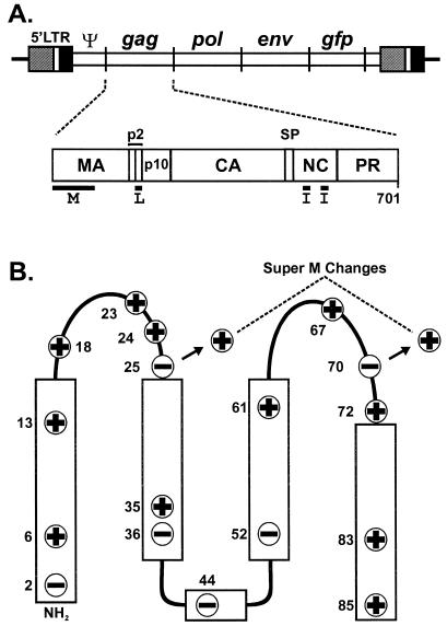
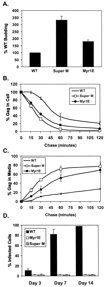
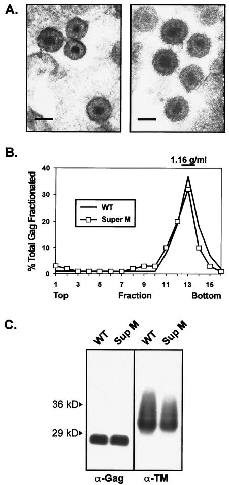
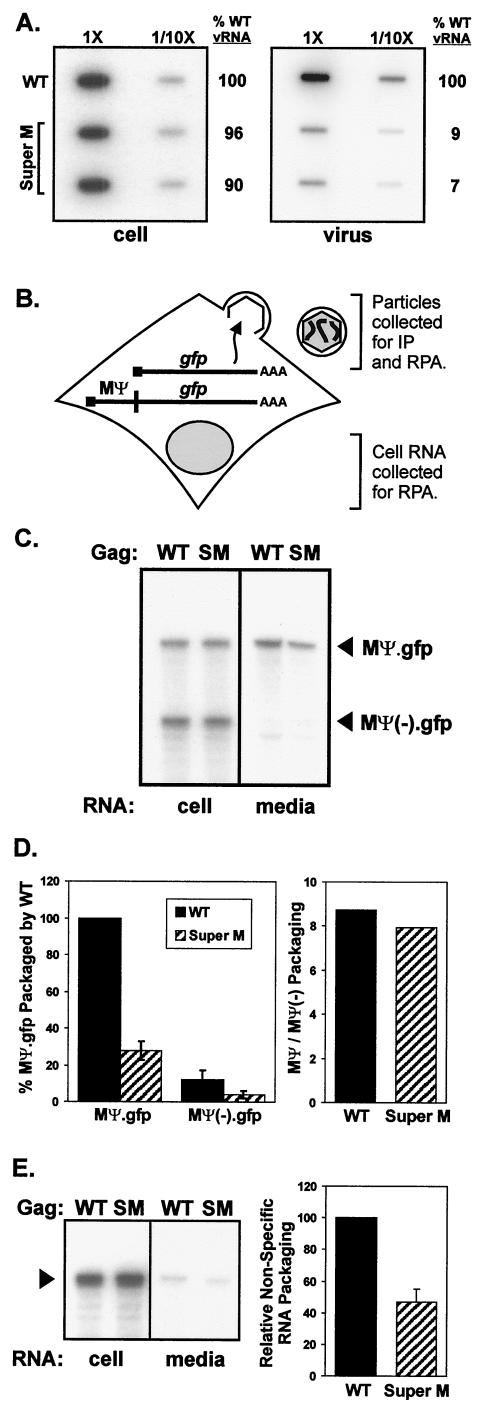
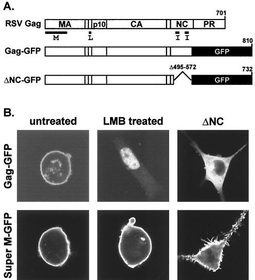
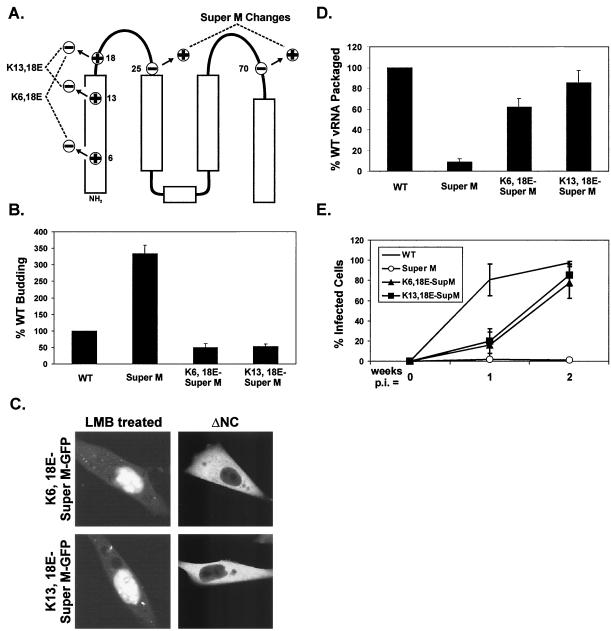
The subcellular location at which genomic RNA is packaged by Gag proteins during retrovirus assembly remains unknown. Since the membrane-binding (M) domain is most critical for targeting Gag to the plasma membrane, changes to this determinant might alter the path taken through the cell and reduce the efficiency of genome packaging. In this report, a Rous sarcoma virus (RSV) mutant having two acidic-to-basic substitutions in the M domain is described. This mutant, designated Super M, produced particles much faster than the wild type, but the mutant virions were noninfectious and contained only 1/10 the amount of genomic RNA found in wild-type particles. To identify the cause(s) of these defects, we considered data that suggest that RSV Gag traffics through the nucleus to package the viral genome. Although inhibition of the CRM-1 pathway of nuclear export caused the accumulation of wild-type Gag in the nucleus, nuclear accumulation did not occur with Super M. The importance of the nucleocapsid (NC) domain in membrane targeting was also determined, and, importantly, deletion of the NC sequence prevented plasma membrane localization by wild-type Gag but not by Super M Gag. Based on these results, we reasoned that the enhanced membrane-targeting properties of Super M inhibit genome packaging. Consistent with this interpretation, substitutions that reestablished the wild-type number of basic and acidic residues in the Super M Gag M domain reduced the budding efficiency and restored genome packaging and infectivity. Therefore, these data suggest that Gag targeting and genome packaging are normally linked to ensure that RSV particles contain viral RNA.


Figures

FIG. 1.
FIG. 1.
Location of the Super M substitutions in RSV Gag. (A) Diagram of the RSV proviral DNA used in these experiments (top) and the RSV Gag polyprotein (bottom). The 5′ long terminal repeat (LTR) promotes transcription of genome-length mRNA, which can be encapsidated or used for the synthesis of Gag and Gag-Pol proteins. In addition, full-length vRNA can be spliced for the synthesis of Env glycoproteins and, in the recombinant virus used here, for the synthesis of GFP. Intact Gag polyproteins drive budding and are subsequently cleaved by the viral PR into the mature products: MA, CA, NC, etc. The locations of the domains required for budding are indicated below Gag. The M domain is essential for plasma membrane targeting. The I domains promote Gag-Gag interactions and the assembly of dense particles. The L domain is required for a late step in budding. (B) The secondary structure of the RSV M domain consists of five helices (rectangles) connected by flexible loops. The locations of the 11 basic and 6 acidic residues in the M domain are depicted according to their charge. The two acidic-to-basic substitutions in Super M Gag, E25K and E70K, are indicated.
FIG. 2.
FIG. 2.
Super M budding and infectivity. (A) QT6 (quail) cells were transfected with proviral (pRCAS-derived) plasmids and labeled for 2.5 h with [35S]methionine. Viral proteins were immunoprecipitated from the cell and media fractions, separated by SDS-PAGE, and quantified by phosphorimager analysis. The amounts of viral proteins in the media were normalized to the levels of intracellular gag expression. The budding efficiency of WT virus was set to 100% for comparison to Super M and the previously described RSV mutant Myr1E, which encodes the Src membrane-binding domain as an extension from the N terminus of Gag. The data are the averages of 10 (Super M) or 4 (Myr1E) experiments, and the error bars measure 1 standard deviation (SD) from the mean. Pulse-chase analyses of Gag levels in the cell (B) and medium (C) fractions were performed with QT6 cells transfected with proviral plasmids. The percentage of Gag in the cells at each time point was determined by dividing the amount remaining by the amount detected at the beginning of the chase. Similarly, the amount of Gag present after the pulse was used to normalize the amount of viral antigen detected in the medium at each time point. The data are the averages of four experiments, and the error bars measure 1 SD from the mean. (D) WT and mutant viruses were collected from transfected QT6 cells, normalized by RT assay, and transferred to cultures of TEFs. Infected cells were detected by FACS analysis because the proviruses expressed gfp from the long terminal repeat. The numbers of infected (fluorescent) cells are expressed as percentages of the total population examined at days 3, 7, and 14 postinfection. The results show the averages of six experiments, and the error bars measure 1 SD from the mean.
FIG. 3.
FIG. 3.
Characterization of Super M particles. (A) Electron micrographs of Super M (left) and WT (right) virus released from QT6 cells transfected with proviral plasmids. (Scale bars = 100 nm). (B) Viral particles were produced by metabolically labeled cells and separated by sucrose density gradient centrifugation. Gag proteins were immunoprecipitated from each gradient fraction, resolved by SDS-PAGE, and quantified by phosphorimager analysis. The amount of Gag in each fraction is expressed as a percentage of the total recovered from all of the fractions collected. WT virus was used as a control for normal density. (C) WT and Super M virions were collected from transfected cells, pelleted through 20% sucrose, and resuspended in PBS. Virus samples corresponding to equivalent amounts of RT activity were denatured, separated by SDS-PAGE, and transferred to nitrocellulose membranes for Western blot analyses. CA bands were detected with polyclonal Gag-reactive antiserum (α-Gag). Env incorporation was determined by using an antibody against the TM subunit (α-TM).
FIG.4.
FIG.4.
Genome packaging properties of Super M. (A) QT6 cells were transfected with proviral plasmids, and particles were prepared as for Fig. 3C. RNA was purified from equivalent numbers of cells and particles, bound to nylon membranes using a slot blot apparatus, and then probed with radiolabeled, antisense RNA specific for the viral genome. Labeled bands were quantified by phosphorimager analysis, and the amounts of vRNA detected in WT cell and virus samples were defined as 100%. To ensure that the RNAs were analyzed within the linear range of this assay, the samples in the right column of each blot contained 1/10 of the total RNA blotted in the left column. (B) Vectors expressing gfp mRNA with or without the RSV minimal packaging sequence (MΨ) were created. These were cotransfected into QT6 cells with WT or Super M gag-only expression vectors (pCMV.GagPR). The cultures were trace labeled with [35S]methionine in 1% serum-containing media. After the labeling, the media were collected and aliquots of each were used to determine the numbers of radiolabeled VLPs present. The remaining media were used for the purification of particle-associated RNA, and the cell-associated RNA was purified from the transfected monolayers. The amounts of MΨ.gfp and MΨ(−).gfp mRNA in both the cell and media fractions were determined by RPA and quantified by phosphorimager analysis. IP, immunoprecipitation. (C) A representative autoradiograph shows the amounts of MΨ.gfp and MΨ(−).gfp mRNA detected in the cell and media fractions by RPA. The specific gag-only expression vector, WT or Super M (SM), cotransfected is indicated above each lane. (D) The packaging efficiencies of MΨ and MΨ(−).gfp mRNA were calculated by dividing the amount detected in the medium by the amount present in the cell. This ratio was then normalized by the number of particles present in the medium. For comparison, the efficiency of MΨ.gfp packaging by WT Gag was set to 100%. The results are the averages of four experiments, and the error bars measure 1 standard deviation (SD) from the mean. In addition, the abilities of WT and Super M Gag proteins to select for Ψ-containing mRNA were calculated by determining the ratio of MΨ to MΨ(−).gfp packaged into extracellular particles. (E) MΨ(−).gfp RNA was coexpressed with either WT or Super M Gag in QT6 cells. RNA was purified from cell and medium fractions, and the amounts of MΨ(−).gfp (arrowhead) present were determined by RPA (left). The relative abilities of WT and Super M Gag to package nonspecific RNA were calculated by normalizing the amount of MΨ(−).gfp RNA in the medium to the amount of Gag released (right). For comparison, the amount of MΨ(−).gfp packaging by WT Gag was set to 100%. The data are the averages of six experiments, and the error bar measures 1 SD from the mean.
FIG. 5.
FIG. 5.
Subcellular localization of Super M Gag. (A) Gag-GFP was created by replacing nonessential, C-terminal sequences of RSV Gag with GFP. The ΔNC-GFP construct contains a large deletion encompassing the I domains in NC. Super M versions of Gag-GFP and ΔNC-GFP were generated by replacing the WT M domain sequences with those with the E25K and E70K substitutions. (B) QT6 cells transfected with Super M or WT Gag-GFP vectors were left untreated or were treated with an inhibitor of nuclear export (LMB) prior to being examined by confocal microscopy. The untreated cells revealed the steady-state localization patterns of WT and Super M Gag-GFP and also served as controls for cells treated with LMB. The ΔNC panels show the effects of deleting NC on the localization of Gag-GFP proteins with WT or Super M domains.
FIG. 6.
FIG. 6.
Creation and analysis of suppressors of Super M. (A) The WT numbers of positively and negatively charged residues were restored in the Super M domain by basic-to-acidic substitutions at positions 6 and 18 or at positions 13 and 18. (B) Analysis of the budding efficiencies of the charge-balanced mutants was performed with proviral vectors as for Fig. 2A. (C) The localization patterns of ΔNC-GFP and Gag-GFP proteins containing charge-balanced M domains in untreated or LMB-treated cells were examined as for Fig. 5B. (D) The genomic RNA content of extracellular virus was determined by using proviral vectors as for Fig. 4A. (E) Infectivity assays were performed at 1 and 2 weeks postinfection as for Fig. 2D. For the results shown in panels B, D, and E, the data are the averages of at least three experiments and the error bars represent 1 standard deviation from the mean.

References

    1. Banks, J. D., A. Yeo, K. Green, F. Cepeda, and M. L. Linial. 1998. A minimal avian retroviral packaging sequence has a complex structure. J. Virol. 72:6190-6194. - PMC - PubMed
    1. Bryant, M., and L. Ratner. 1990. Myristoylation-dependent replication and assembly of human immunodeficiency virus 1. Proc. Natl. Acad. Sci. USA 87:523-527. - PMC - PubMed
    1. Burniston, M. T., A. Cimarelli, J. Colgan, S. P. Curtis, and J. Luban. 1999. Human immunodeficiency virus type 1 Gag polyprotein multimerization requires the nucleocapsid domain and RNA and is promoted by the capsid-dimer interface and the basic region of matrix protein. J. Virol. 73:8527-8540. - PMC - PubMed
    1. Callahan, E. M., and J. W. Wills. 2000. Repositioning basic residues in the M domain of the Rous sarcoma virus Gag protein. J. Virol. 74:11222-11229. - PMC - PubMed
    1. Campbell, S., and V. M. Vogt. 1995. Self-assembly in vitro of purified CA-NC proteins from Rous sarcoma virus and human immunodeficiency virus type 1. J. Virol. 69:6487-6497. - PMC - PubMed

Publication types

LinkOut - more resources