. 2003 Sep;77(17):9502-10.
doi: 10.1128/jvi.77.17.9502-9510.2003.

Complementation of a deletion in the rubella virus p150 nonstructural protein by the viral capsid protein

Affiliations

Complementation of a deletion in the rubella virus p150 nonstructural protein by the viral capsid protein

Wen-Pin Tzeng et al. J Virol. 2003 Sep.

Abstract

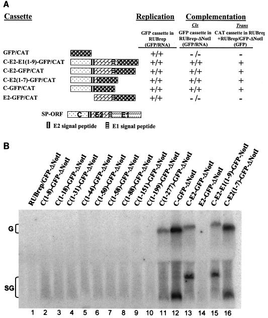
Rubella virus (RUB) replicons with an in-frame deletion of 507 nucleotides between two NotI sites in the P150 nonstructural protein (DeltaNotI) do not replicate (as detected by expression of a reporter gene encoded by the replicon) but can be amplified by wild-type helper virus (Tzeng et al., Virology 289:63-73, 2001). Surprisingly, virus with DeltaNotI was viable, and it was hypothesized that this was due to complementation of the NotI deletion by one of the virion structural protein genes. Introduction of the capsid (C) protein gene into DeltaNotI-containing replicons as an in-frame fusion with a reporter gene or cotransfection with both DeltaNotI replicons and RUB replicon or plasmid constructs containing the C gene resulted in replication of the DeltaNotI replicon, confirming the hypothesis that the C gene was the structural protein gene responsible for complementation and demonstrating that complementation could occur either in cis or in trans. Approximately the 5' one-third of the C gene was necessary for complementation. Mutations that prevented translation of the C protein while minimally disturbing the C gene sequence abrogated complementation, while synonymous codon mutations that changed the C gene sequence without affecting the amino acid sequence at the 5' end of the C gene had no effect on complementation, indicating that the C protein, not the C gene RNA, was the moiety responsible for complementation. Complementation occurred at a basic step in the virus replication cycle, because DeltaNotI replicons failed to accumulate detectable virus-specific RNA.


Figures

FIG. 1.
FIG. 1.
Mapping the moiety responsible for complementation. In panel A and Table 1 are shown the results of experiments with cassettes expressed by RUBrep or RUBrep-ΔNotI. The cassettes consisted of either a reporter gene (GFP or CAT) or a portion of the RUB or SIN SP-ORF fused in frame with GFP or CAT. The GFP version of each cassette was used to assess replication of the wild-type replicon, RUBrep (replication column), and of the replicon with the NotI deletion, RUBrep-ΔNotI (cis complementation column), containing the cassette. Replication or complementation was detected by both GFP expression using fluorescence microscopy of transfected cells (GFP) and virus-specific RNA production using Northern blot analysis (B [RNA]). trans complementation was assessed by cotransfecting cells with transcripts of the RUBrep construct containing the CAT version of the cassette and RUBrep/GFP-ΔNotI transcripts; replication of RUBrep/GFP-ΔNotI was detected by GFP expression. Panel A summarizes experiments with fusion proteins containing portions of the RUB SP-ORF. A schematic diagram of the ORF with the coding sequences for the C, E2, and E1 proteins as well as the E2 and E1 signal sequences (which remain attached to C and E2, respectively, following processing) is shown at the top of the panel and the portion of the ORF contained in each cassette, along with the reporter gene, is shown under this diagram. The C-E2-E1(1-9)-GFP/CAT cassette contains the complete C and E2 genes and the first nine amino acids of the E1 gene fused to the reporter gene, and the C-E2(1-7)-GFP/CAT cassette contains the complete C gene and the first seven amino acids of the E2 gene fused to the reporter gene. For the Northern blot shown in panel B, Vero cells were transfected with transcripts from RUBrep/GFP-ΔNotI (lane 1), RUBrep-ΔNotI constructs expressing a series of C-GFP-ΔNotI fusions that contained progressive 3′-terminal deletions of the C gene (lanes 2 to 11), RUBrep/C-GFP-ΔNotI (lane 12), RUBrep/C-E2-GFP-ΔNotI (lane 13), RUBrep/E2-GFP-ΔNotI (lane 14), RUBrep/C-E2-E1(1-9)-GFP-ΔNotI (lane 15), or RUBrep/C-E2(1-7)-GFP-ΔNotI (lane 16). Four days posttransfection, total RNA was extracted and analyzed by gel electrophoresis, blotting, and probing with 32P-labeled pGEM-GFP DNA. The positions of migration of the replicon genomic (G) RNAs and SG RNAs (which vary in size) are denoted.
FIG. 2.
FIG. 2.
Viability and trans complementation by infectious cDNA clone constructs. Shown are genomic diagrams of Robo502 and Robo502/IRES, in which the junction-UTR between the NS- and SP-ORFs was replaced by the IRES of encephalomycarditis virus, without and with the NotI deletion in the P150 gene. As shown, all of these constructs give rise to viable virus, as shown by the titers and plaque morphologies of virus in transfection culture fluid harvested 4 days posttransfection. To test for the ability to complement the NotI deletion in trans, Vero cells were cotransfected with transcripts from one of these constructs and RUBrep/GFP-ΔNotI transcripts, and replication of RUBrep/GFP-ΔNotI was detected by GFP expression.
FIG. 3.
FIG. 3.
Effect on complementation of mutations at the 5′ end of the C gene. Using the C-GFP/CAT cassette, a series of mutations at the 5′ end of the C gene were made. (The 5′ end of the C gene has two in-frame AUGs separated by seven codons). Under the wild-type sequence, each mutated sequence is given with the mutation in boldface and designated with an asterisk. Each mutated cassette was assessed for replication in wild-type replicon RUBrep/GFP, cis complementation in RUBrep/GFP-ΔNotI, and trans complementation in RUBrep/CAT (in cells cotransfected with RUBrep/GFP-ΔNotI transcripts) as described in the legend to Fig. 1 and shown in Table 1.
FIG. 4.
FIG. 4.
Alignment of the sequences of RUB C protein with those of the SIN C protein and NotI domain. The entire RUB and SIN C protein sequences are included in the alignment; amino acids are numbered from the N-terminal Met residue. In the RUB C X NotI alignment, only the N-terminal 100 residues of the C protein are used. The numbering of the NotI domain represents the residue number within the P150 protein. The alignment was made with the MacVector version 6.5.3 program.

Similar articles

Cited by

References

    1. Adams, S. D., W.-P. Tzeng, M.-H. Chen, and T. K. Frey. 2003. Analysis of intermolecular RNA-RNA recombination by rubella virus. Virology 309:258-271. - PMC - PubMed
    1. Barry, J. K., and W. A. Miller. 2002. A −1 ribosomal frameshift element that requires base pairing across four kilobases suggests a mechanism of regulating ribosome and replicase traffic on a viral RNA. Proc. Natl. Acad. Sci. USA 99:11133-11138. - PMC - PubMed
    1. Beatch, M. D., and T. C. Hobman. 2000. Rubella virus capsid associates with host cell protein p32 and localizes to mitochondria. J. Virol. 74:5569-5576. - PMC - PubMed
    1. Chen, J.-P., J. H. Strauss, E. G. Strauss, and T. K. Frey. 1996. Characterization of the rubella virus nonstructural protease domain and its cleavage site. J. Virol. 70:4707-4713. - PMC - PubMed
    1. Choi, I. R., and K. A. White. 2002. An RNA activator of subgenomic mRNA1 transcription in tomato bushy stunt virus. J. Biol. Chem. 277:3760-3766. - PubMed

Publication types

MeSH terms

LinkOut - more resources