. 2003 Sep;77(17):9511-21.
doi: 10.1128/jvi.77.17.9511-9521.2003.

Pre-s1 antigen-dependent infection of Tupaia hepatocyte cultures with human hepatitis B virus

Affiliations

Pre-s1 antigen-dependent infection of Tupaia hepatocyte cultures with human hepatitis B virus

Dieter Glebe et al. J Virol. 2003 Sep.

Erratum in

  • J Virol. 2005 Nov;79(22):14470

Abstract

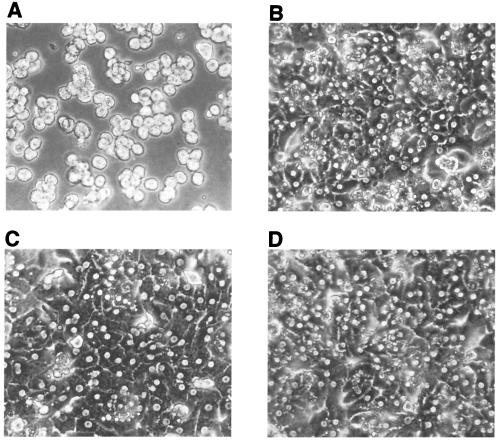
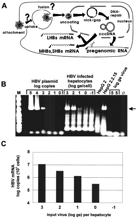
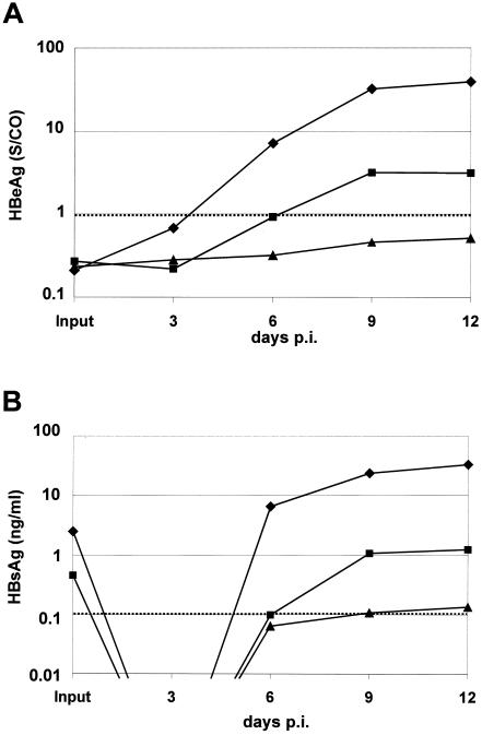
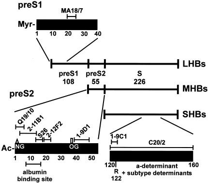
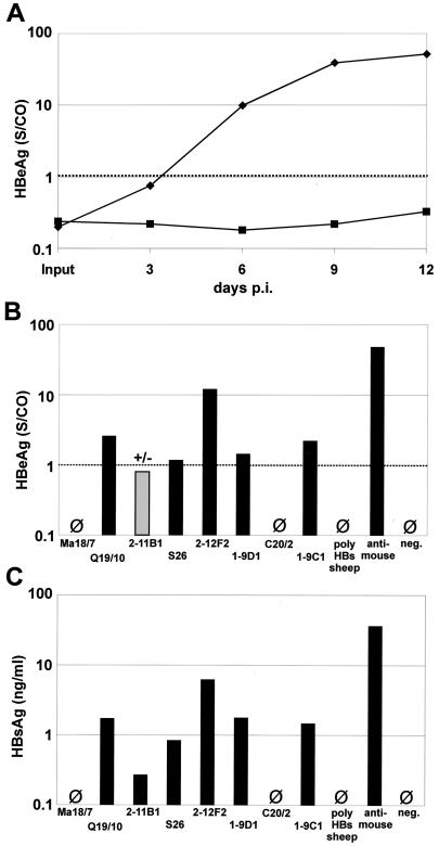
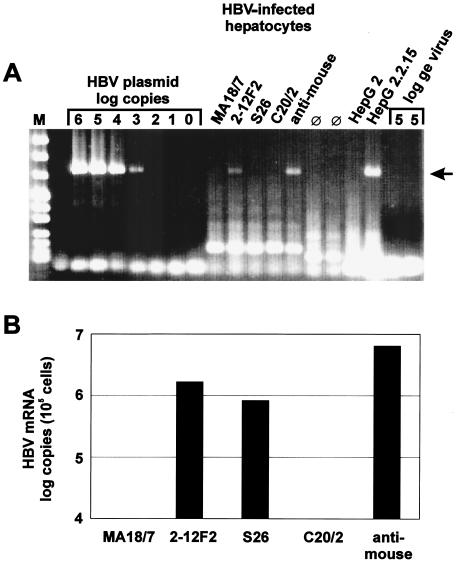
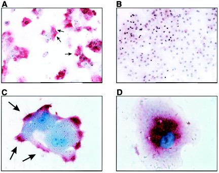
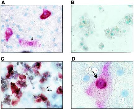
The susceptibility of the tree shrew Tupaia belangeri to human hepatitis B virus (HBV) has been demonstrated both in vivo and in vitro. In this study, we show that purified HBV infects primary T. belangeri hepatocyte cultures in a very specific manner, as detected by HBV covalently closed circular DNA, mRNA, HBV e antigen, and HBsAg production. A monoclonal antibody (MAb), MA18/7, directed against the pre-S1 domain of the large HBs protein, which has been shown to neutralize infectivity of HBV for primary human hepatocytes, also blocked infection of primary Tupaia hepatocytes. MAbs against the pre-S2 domain of HBs inhibited infection only partially, whereas an S MAb and polyvalent anti-HBs antibodies neutralized infection completely. Thus, both pre-S1 and S antigens are necessary for infection in the tupaia. Using subviral particles, >70% of primary Tupaia hepatocytes are capable of specific binding of pre-S1-rich HBsAg, showing localization in distinct membrane areas. The data show that the early steps of HBV infection in Tupaia hepatocyte cultures are comparable to those in the human system.


Figures

FIG. 1.
FIG. 1.
Morphologies of primary Tupaia hepatocyte cultures. Shown are phase-contrast micrographs of uninfected primary Tupaia hepatocytes at various time points after two-step collagenase isolation. (A) After perfusion; (B) day 3; (C) day 6; (D) day 9. Hepatocytes preserve their shape and functionality during the time required for infection. Note the frequent presence of two nuclei and the bright borders between the cells (bile canaliculi), typical features of well-differentiated hepatocytes (magnification, ×100).
FIG. 2.
FIG. 2.
Detection of replicative intermediates in primary Tupaia hepatocytes infected with increasing amounts of purified virus. (A) schematic diagram of the early events in HBV infection of hepatocytes. Formation of cccDNA from the incoming HBV rcDNA form with its nick-and-gap structure is essential for RNA transcription and all further steps in establishing an HBV infection. (B) Agarose gel electrophoresis of PCR products specific for HBV double-stranded DNA without nick and gap. A specific amplificate at 735 bp (arrow) could be synthesized from cloned dimeric HBV DNA but not from purified virion rcDNA, with its nick-and-gap structure, from human plasma. HBV cccDNA was detected in nuclear Hirt extracts of HBV-infected primary Tupaia hepatocytes 12 days p.i. or in the positive control cell line HepG2.2.15, but not from the parental HepG2 cell line. Lane M, marker; lane ⊘, negative control. (C) HBV mRNA quantification of cytoplasmic extracts from primary Tupaia hepatocytes 12 days after infection with increasing amounts of purified HBV (real-time RT-PCR of the X-gene region). The detection limit was 104 copies per culture. ge, genomic equivalent.
FIG. 3.
FIG. 3.
HBeAg (A) and HBsAg (B) secretion of primary Tupaia hepatocytes infected with increasing amounts of purified HBV. The medium was changed every 3 days. Each point represents HBeAg or HBsAg in the supernatant newly synthesized within 3 days. ⧫, 100 ge/cell; ▪, 10 ge/cell; ▴, 1 ge/cell. The dotted lines indicate the cutoffs for HBeAg (multiples of cutoff signal) and for HBsAg (0.1 ng/ml). S/CO, sample to cutoff signal.
FIG. 4.
FIG. 4.
Neutralizing epitopes of antibodies against HBV surface proteins. Schematic diagram of MAb epitopes of LHBs, MHBs, and SHBs (genotype D). NG, N-glycan in the pre-S2 domain of MHBs; OG, O-linked glycan. R122 refers to the subtype determinant y in the antigenic region of SHBs.
FIG. 5.
FIG. 5.
Neutralization test using HBeAg or HBsAg secretion as readout. (A) Kinetic analysis of HBeAg production of infected primary Tupaia hepatocytes after preincubation of input virus with anti-pre-S1 MAb MA18/7 (⧫) or with an irrelevant anti-mouse MAb (▪) (the dotted line indicates the cutoff). (B) HBeAg production of infected primary Tupaia hepatocytes 12 days p.i. after preincubation of input virus with different MAbs against HBV surface proteins. (C) HBsAg production of infected primary Tupaia hepatocytes 12 days p.i. after preincubation of input virus with different MAbs against HBV surface proteins (the cutoff for HBsAg was 0.1 ng/ml).
FIG. 6.
FIG. 6.
Inhibition of HBV cccDNA formation and transcription by pretreatment of HBV with neutralizing MAbs. (A) Agarose gel electrophoresis of PCR products specific for HBV DNA double stranded in the nick-gap region as described in the legend to Fig. 2B. The purified virus inoculum was preincubated before infection with MAbs Ma18/7 (pre-S1), 2-12F2 (pre-S2), S26 (pre-S2), and C20/2 (S) and an irrelevant anti-mouse antibody (anti-mouse). Lane M, marker; lane ⊘, negative control. (B) HBV mRNA quantification of cytoplasmic extracts from HBV-infected primary Tupaia hepatocytes 12 days p.i. by real-time RT-PCR. The purified input virus was preincubated before infection with MAbs as for panel A.
FIG. 7.
FIG. 7.
Binding and uptake of subviral HBsAg particles. Shown is immune staining of Tupaia hepatocytes after incubation with purified LHBs-rich HBV subviral particles for 1 h at 37°C with a MAb against the S domain (red) and counterstaining with Meyer's hemalaun (blue). (A, C, and D) HBsAG staining; (C) inhibition by polyclonal anti-HBs. (A and B) Binding of HBsAg particles to primary Tupaia hepatocytes is detected (A) (arrows) (magnification, ×100), but not after preincubation of the inoculum with polyclonal anti-HBs antibodies (B) (magnification, ×100). (C) At higher magnification, HBs particles (arrows) are detected in distinct areas of the plasma membrane (magnification, ×400). (D) Some hepatocytes show a pattern suggesting uptake of HBsAg (magnification, ×630).
FIG. 8.
FIG. 8.
(A, C, and D) HBsAg immune staining of infected Tupaia hepatocytes. Tupaia hepatocytes were infected with purified HBV (100 ge/cell) as described in Materials and Methods and immune stained for newly produced HBsAg 12 days p.i. (A) (magnification, ×200). Infection is inhibited by preincubation of virus inoculum with polyclonal anti-HBs antibody (B) (magnification ×200). Increased viral input (10,000 ge/hepatocyte) resulted in infection of up to 20% of hepatocytes (C) (magnification, ×200). HBsAg is distributed within the cytoplasm (D) (magnification, ×630) with typical perinuclear accumulation (arrow).

Similar articles

Cited by

References

    1. Aldrich, C. E., L. Coates, T. T. Wu, J. Newbold, B. C. Tennant, J. Summers, C. Seeger, and W. S. Mason. 1989. In vitro infection of woodchuck hepatocytes with woodchuck hepatitis virus and ground squirrel hepatitis virus. Virology 172:247-252. - PubMed
    1. Bruss, V., X. Lu, R. Thomssen, and W. H. Gerlich. 1994. Post-translational alterations in transmembrane topology of the hepatitis B virus large envelope protein. EMBO J. 13:2273-2279. - PMC - PubMed
    1. Chouteau, P., J. Le Seyec, I. Cannie, M. Nassal, C. Guguen-Guillouzo, and P. Gripon. 2001. A short N-proximal region in the large envelope protein harbors a determinant that contributes to the species specificity of human hepatitis B virus. J. Virol. 75:11565-11572. - PMC - PubMed
    1. Cooreman, M. P., G. Leroux-Roels, and W. P. Paulij. 2001. Vaccine- and hepatitis B immune globulin-induced escape mutations of hepatitis B virus surface antigen. J. Biomed. Sci. 8:237-247. - PubMed
    1. Dandri, M., M. R. Burda, E. Torok, J. M. Pollok, A. Iwanska, G. Sommer, X. Rogiers, C. E. Rogler, S. Gupta, H. Will, H. Greten, and J. Petersen. 2001. Repopulation of mouse liver with human hepatocytes and in vivo infection with hepatitis B virus. Hepatology 33:981-988. - PubMed

Publication types

Substances

LinkOut - more resources