. 2003 Sep;77(17):9522-32.
doi: 10.1128/jvi.77.17.9522-9532.2003.

Newcastle disease virus V protein is a determinant of host range restriction

Affiliations

Newcastle disease virus V protein is a determinant of host range restriction

Man-Seong Park et al. J Virol. 2003 Sep.

Abstract

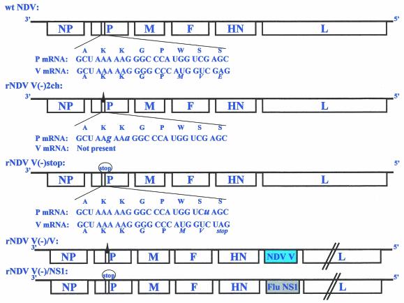
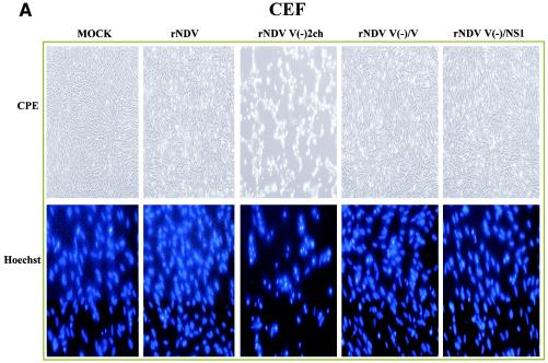
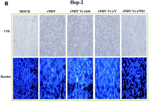
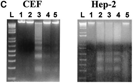
It has been demonstrated that the V protein of Newcastle disease virus (NDV) functions as an alpha/beta interferon (IFN-alpha/beta) antagonist (M. S. Park, M. L. Shaw, J. Muñoz-Jordan, J. F. Cros, T. Nakaya, N. Bouvier, P. Palese, A. García-Sastre, and C. F. Basler, J. Virol. 77:1501-1511, 2003). We now show that the NDV V protein plays an important role in host range restriction. In order to study V functions in vivo, recombinant NDV (rNDV) mutants, defective in the expression of the V protein, were generated. These rNDV mutants grow poorly in both embryonated chicken eggs and chicken embryo fibroblasts (CEFs) compared to the wild-type (wt) rNDV. However, insertion of the NS1 gene of influenza virus A/PR8/34 into the NDV V(-) genome [rNDV V(-)/NS1] restores impaired growth to wt levels in embryonated chicken eggs and CEFs. These data indicate that for viruses infecting avian cells, the NDV V protein and the influenza NS1 protein are functionally interchangeable, even though there are no sequence similarities between the two proteins. Interestingly, in human cells, the titer of wt rNDV is 10 times lower than that of rNDV V(-)/NS1. Correspondingly, the level of IFN secreted by human cells infected with wt rNDV is much higher than that secreted by cells infected with the NS1-expressing rNDV. This suggests that the IFN antagonist activity of the NDV V protein is species specific. Finally, the NDV V protein plays an important role in preventing apoptosis in a species-specific manner. The rNDV defective in V induces apoptotic cell death more rapidly in CEFs than does wt rNDV. Taken together, these data suggest that the host range of NDV is limited by the ability of its V protein to efficiently prevent innate host defenses, such as the IFN response and apoptosis.


Figures

FIG. 1.
FIG. 1.
Schematic representation of rNDV mutants. The P and V mRNAs expressed from wt NDV are indicated at the top. The P and V mRNAs are identical except for one nontemplated G nucleotide in the V mRNA. The amino acids encoded by the indicated nucleotide sequences are also shown. Amino acids in italics show coding differences in the P and V proteins due to mRNA editing. Mutations introduced into rNDV V(−)2ch are indicated by a solid triangle. The mutation introduced into rNDV V(−)stop is also indicated by a circled “stop.” rNDV V(−)2ch is deficient in expression of V and W proteins due to an impairment in RNA editing. To selectively block the expression of the carboxy terminus of the V protein, a stop codon was created in the V ORF without affecting the P frame in rNDV V(−)stop. The NDV V or the influenza virus (Flu) PR8 NS1 gene was inserted between the HN and L genes in the context of rNDV V(−)2ch or rNDV V(−)stop, respectively, to generate rNDV V(−)/V and rNDV V(−)/NS1. For details, see Materials and Methods.
FIG. 2.
FIG. 2.
Growth kinetics of rNDV mutants. CEFs and Vero, Hep-2, and A549 cells were infected with each indicated virus at an MOI of 0.01 in the presence of 10% allantoic fluid. In addition, 10-day-old embryonated chicken eggs were inoculated with 100 TCID50/egg. Each supernatant from infected cells and embryonated eggs was harvested every 24 h for 3 days. Titers were determined by measuring the TCID50 in Vero cells. Note that the mutations responsible for the loss of the V function in rNDV V(−)/V and rNDV V(−)/NS1 are due to the insertion of an editing mutation and a stop codon, respectively.
FIG. 3.
FIG. 3.
Expression of the influenza A virus NS1 protein by rNDV V(−)/NS1. Expression of the influenza virus NS1 protein or of NDV viral proteins was detected in infected CEFs by immunofluorescence using anti-rabbit influenza virus NS1 polyclonal antibody or anti-rabbit NDV polyclonal antibody, respectively. Magnification, ×10.
FIG. 4.
FIG. 4.
IFN production by rNDV mutant-infected cells. To measure IFN production after viral infection, cells were infected with rNDV or rNDV mutants at an MOI of 1. Supernatants (Sup) from infected cells were harvested 24 h p.i. and then treated with UV light for 7 min to inactivate the infectivity of the virus. UV-treated supernatants were added to new Vero cells or CEFs. The UV-treated supernatants were removed 20 h posttreatment, and then the cells were infected with rNDV-GFP at an MOI of 1. NDV-GFP expression was analyzed by fluorescence microscopy 24 h p.i. Note that the mutations responsible for the loss of the V function in rNDV V(−)/V and rNDV V(−)/NS1 are due to the insertion of an editing mutation and a stop codon, respectively.
FIG.5
FIG.5
Induction of apoptosis by rNDV mutants. CEFs and Hep-2 cells were infected with wt rNDV or rNDV mutants at an MOI of 1 or mock infected. (A and B) The morphologies of infected cells at 36 h p.i. were analyzed by phase-contrast microscopy (CPE; magnification, 310). To observe chromatin condensation in cell nuclei, the infected cells at 36 h p.i. were fixed with methanol containing 10% formaldehyde and stained with the DNA-binding dye Hoechst 33258 (magnification, 340). (C) DNA fragmentation was analyzed 36 h p.i. Lane 1, uninfected cells; lane 2, wt rNDV-infected cells (MOI 5 1); lane 3, rNDV V(2)2ch-infected cells (MOI 5 1); lane 4, rNDV V(2)/V-infected cells (MOI 5 1); lane 5, rNDV V(2)/NS1-infected cells (MOI 5 1); L, 1-kb DNA ladder. (D) TUNEL staining of rNDV-infected CEFs and Hep-2 cells at 24 and 48 h p.i. Note that the mutations responsible for the loss of the V function in rNDV V(2)/V and rNDV V(2)/NS1 are due to the insertion of an editing mutation and a stop codon, respectively.—Continued.
FIG.5
FIG.5
Induction of apoptosis by rNDV mutants. CEFs and Hep-2 cells were infected with wt rNDV or rNDV mutants at an MOI of 1 or mock infected. (A and B) The morphologies of infected cells at 36 h p.i. were analyzed by phase-contrast microscopy (CPE; magnification, 310). To observe chromatin condensation in cell nuclei, the infected cells at 36 h p.i. were fixed with methanol containing 10% formaldehyde and stained with the DNA-binding dye Hoechst 33258 (magnification, 340). (C) DNA fragmentation was analyzed 36 h p.i. Lane 1, uninfected cells; lane 2, wt rNDV-infected cells (MOI 5 1); lane 3, rNDV V(2)2ch-infected cells (MOI 5 1); lane 4, rNDV V(2)/V-infected cells (MOI 5 1); lane 5, rNDV V(2)/NS1-infected cells (MOI 5 1); L, 1-kb DNA ladder. (D) TUNEL staining of rNDV-infected CEFs and Hep-2 cells at 24 and 48 h p.i. Note that the mutations responsible for the loss of the V function in rNDV V(2)/V and rNDV V(2)/NS1 are due to the insertion of an editing mutation and a stop codon, respectively.—Continued.

Similar articles

Cited by

References

    1. Basler, C., A. Reid, J. Dybing, T. Janczewski, T. Fanning, H. Zheng, M. Salvatore, M. Perdue, D. Swayne, A. Garcia-Sastre, P. Palese, and J. Taubenberger. 2001. Sequence of the 1918 pandemic influenza virus nonstructural gene (NS) segment and characterization of recombinant viruses bearing the 1918 NS genes. Proc. Natl. Acad. Sci. USA 98:2746-2751. - PMC - PubMed
    1. Beard, C. W., and R. P. Hanson. 1984. Newcastle disease, p. 452-470. In M. S. Hofstad, H. J. Barnes, B. W. Calnek, W. M. Reid, and W. Yoder (ed.), Disease of poultry. Iowa State University Press, Ames, Iowa.
    1. Bossert, B., and K. K. Conzelmann. 2002. Respiratory syncytial virus (RSV) nonstructural (NS) proteins as host range determinants: a chimeric bovine RSV with NS genes from human RSV is attenuated in interferon-competent bovine cells. J. Virol. 76:4287-4293. - PMC - PubMed
    1. Didcock, L., D. F. Young, S. Goodbourn, and R. E. Randall. 1999. The V protein of simian virus 5 inhibits interferon signalling by targeting STAT1 for proteasome-mediated degradation. J. Virol. 73:9928-9933. - PMC - PubMed
    1. Garcia-Sastre, A. 2001. Inhibition of interferon-mediated antiviral responses by influenza A viruses and other negative-strand RNA viruses. Virology 279:375-384. - PubMed

Publication types

MeSH terms

LinkOut - more resources