. 2003 Sep;77(17):9542-52.
doi: 10.1128/jvi.77.17.9542-9552.2003.

Specific association of glycoprotein B with lipid rafts during herpes simplex virus entry

Affiliations

Specific association of glycoprotein B with lipid rafts during herpes simplex virus entry

Florent C Bender et al. J Virol. 2003 Sep.

Abstract

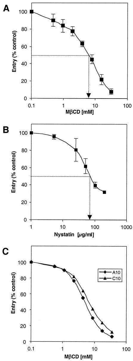
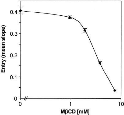
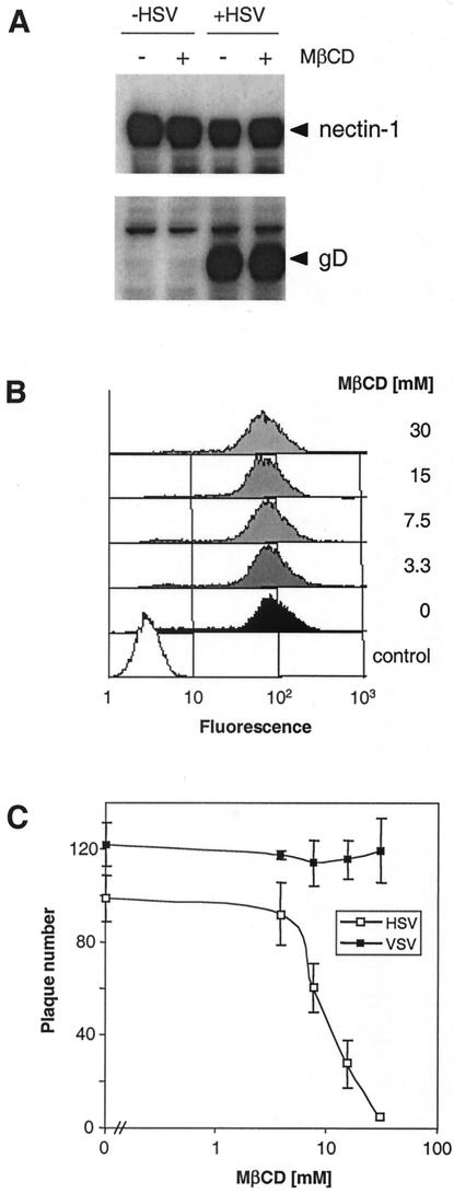
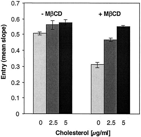
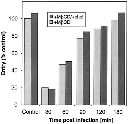
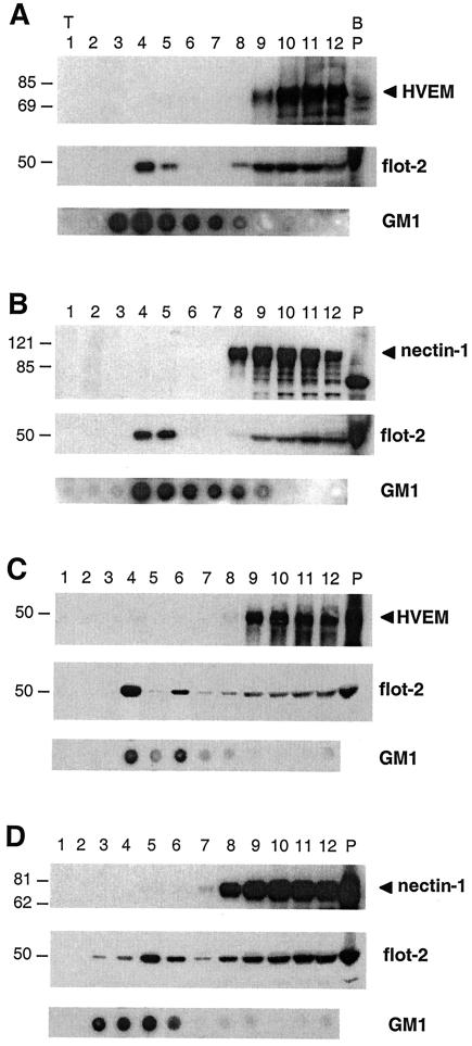
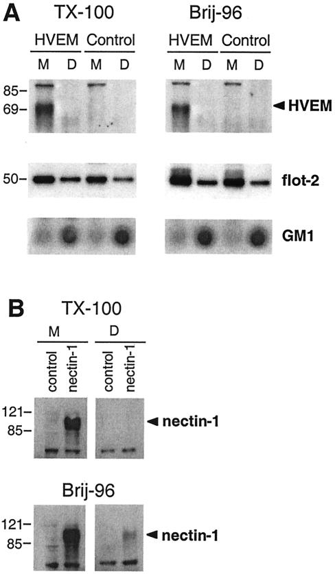
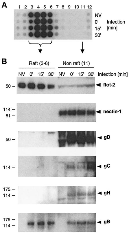
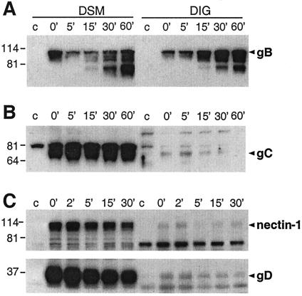
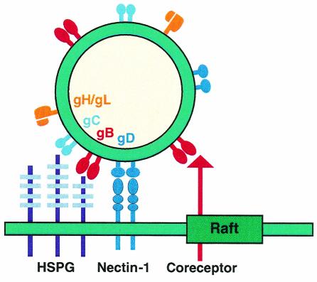
Herpes simplex virus (HSV) entry requires the interaction of glycoprotein D (gD) with a cellular receptor such as herpesvirus entry mediator (HVEM or HveA) or nectin-1 (HveC). However, the fusion mechanism is still not understood. Since cholesterol-enriched cell membrane lipid rafts are involved in the entry of other enveloped viruses such as human immunodeficiency virus and Ebola virus, we tested whether HSV entry proceeds similarly. Vero cells and cells expressing either HVEM or nectin-1 were treated with cholesterol-sequestering drugs such as methyl-beta-cyclodextrin or nystatin and then exposed to virus. In all cases, virus entry was inhibited in a dose-dependent manner, and the inhibitory effect was fully reversible by replenishment of cholesterol. To examine the association of HVEM and nectin-1 with lipid rafts, we analyzed whether they partitioned into nonionic detergent-insoluble glycolipid-enriched membranes (DIG). There was no constitutive association of either receptor with DIG. Binding of soluble gD or virus to cells did not result in association of nectin-1 with the raft-containing fractions. However, during infection, a fraction of gB but not gC, gD, or gH associated with DIG. Similarly, when cells were incubated with truncated soluble glycoproteins, soluble gB but not gC was found associated with DIG. Together, these data favor a model in which HSV uses gB to rapidly mobilize lipid rafts that may serve as a platform for entry and cell signaling. It also suggests that gB may interact with a cellular molecule associated with lipid rafts.


Figures

FIG. 1.
FIG. 1.
Effect of cholesterol depletion or chelation on HSV entry. Vero cells (A and B) and B78 A10 and C10 cells (C) were treated with increasing concentrations of MβCD (A and C) or nystatin (B). The drugs were washed out, and the cells were infected with HSV-1 (KOS/tk12) and assayed for β-galactosidase activity at 6 h postinfection. Results presented in A and B are the mean of three independent experiments done in duplicate, with standard deviations. The results shown in panel C are representative of three experiments. A value of 100% entry represents the β-galactosidase activity at 6 h in the absence of MβCD.
FIG. 2.
FIG. 2.
Effect of cholesterol depletion from virions on HSV entry. HSV-1 (KOS/tk12) particles (2 × 10⁶ PFU) were treated with various amounts of MβCD for 30 min at room temperature. MβCD was then diluted to a final concentration of 0.1 mM or less, a concentration that has no effect on cells, and virus was allowed to infect Vero cells (multiplicity of infection of 10). β-Galactosidase activity at 6 h postinfection was determined as described for Fig. 1. The results presented are representative of two independent experiments done in triplicate, with standard deviations.
FIG. 3.
FIG. 3.
Effect of cholesterol depletion from cells on expression of nectin-1 and on attachment and replication of HSV. (A) C10 cells were untreated (−) or treated (+) with 30 mM MβCD for 30 min. The drug was washed out, and HSV-1 (KOS) was allowed to bind to the cells for 1 h at 4°C. Total cell proteins were extracted, resolved by SDS-PAGE, and probed with anti-nectin-1 MAb CK6 (upper panel) or anti-gD MAb DL6 (lower panel). (B) C10 cells were treated with increasing concentrations of MβCD (indicated at the right), then stained with anti-nectin-1 MAb CK41 directly labeled with phycoerythrin, and analyzed by FACS. The control (open area) represents the fluorescence of unstained and drug untreated cells. (C) Vero cells were treated with increasing concentrations of MβCD. The drug was washed out, and the cells were infected with HSV1 (KOS) or vesicular stomatitis virus (VSV) for 24 h at 37°C. Then the cells were fixed, and plaques werevisualized by immunoperoxidase staining (HSV) or crystal violet staining (vesicular stomatitis virus).
FIG. 4.
FIG. 4.
Effect of cholesterol replenishment on HSV entry. Vero cells were untreated (left, rectangles) or treated (right, rectangles) with 7.5 mM MβCD for 30 min. MβCD was washed out, and various amounts of cholesterol in MβCD were added. After 30 min, the cholesterol was removed, and the cells were infected with HSV-1 (KOS/tk12) and assayed for β-galactosidase activity at 6 h postinfection. The results presented are representative of two independent experiments done in triplicate.
FIG. 5.
FIG. 5.
Effect of cholesterol depletion from cells on the course of HSV entry. Vero cells were infected with HSV-1 (KOS/tk12). At various times following infection (indicated on the abscissa), cells were treated for 30 min with MβCD. The drug was washed out, and medium or cholesterol (chol) was added. After 30 min, the cholesterol was removed, and infection was allowed to proceed. β-Galactosidase activity at 6 h postinfection was determined. The results shown are representative of three independent experiments, and 100% entry represents the β-galactosidase activity at 6 h in the absence of MβCD.
FIG. 6.
FIG. 6.
Association of HSV receptors with detergent-insoluble low-density sucrose fractions containing rafts. 3E5 cells stably expressing HVEM-GFP (A), C10 cells stably expressing nectin-1 (B), and 293T cells transiently transfected with a plasmid expressing the HVEM (C) or nectin-1 (D) gene were homogenized in Triton X-100 and fractionated on sucrose gradients. Proteins (10 μl of each fraction) were resolved on SDS-PAGE and transferred to nitrocellulose, and the distribution of HSV receptors was analyzed by Western blotting with MAb CW10 to HVEM or MAb CK6 to nectin-1. Fraction numbers are indicated at the top of the figure (P for pellet). T and B refer to the top and bottom of the gradient, respectively. The distribution of the known raft-associated protein flotillin-2 (flot-2) was analyzed with a commercial MAb and served as a positive control for rafts (A, B, C, and D, middle panels). Also shown is the distribution of the raft-specific ganglioside GM1 analyzed by dot blot with CTB-HRP (A, B, C, and D, lower panels). Sizes are shown on the left (in kilodaltons).
FIG. 7.
FIG. 7.
Association of HSV receptors with detergent-insoluble glycolipid complexes. 3E5 cells (A), C10 cells (B), and control parental B78-H1 cells were fractionated into detergent-soluble (DSM) and insoluble (DIG) membrane fractions with either Triton X-100 (TX-100) or Brij-96. The distribution of the two HSV receptors was analyzed by Western blotting as described for Fig. 6. At the top are indicated the receptor expressed (control, HVEM, or nectin-1), the detergent used (Triton X-100 or Brij-96), and the fraction, DSM (M) or DIG (D). Sizes are shown on the left (in kilodaltons).
FIG. 8.
FIG. 8.
Association of HSV receptors and glycoproteins with detergent-insoluble low-density sucrose fractions during entry. C10 cells were infected with HSV (KOS), and at various times postinfection, cells were extracted in Triton X-100 and fractionated on a sucrose gradient as in Fig. 6. (A) The distribution of GM1 in the different sucrose gradient fractions was analyzed by dot blot in noninfected cells (NV) and after infection for various times, indicated at the right of the panel. (B) Low-density fractions 3 to 6 containing the raft marker GM1 (raft) were pooled, precipitated with methanol-chloroform, resuspended in gel sample buffer, and resolved by SDS-PAGE. The distribution of flotillin-2 (flot-2) and nectin-1 at various times during infection (indicated at the top of the panel) was compared to that found in 10 μl of the GM1-negative fraction 11 (non-raft) by Western blotting on separate membranes. The distribution of the glycoproteins (indicated at the right) in raft and nonraft fractions was analyzed with MAb DL6 to gD, polyclonal antibody R47 to gC, polyclonal antibody R137 to gH, and MAb SS-10 to gB. Sizes are shown on the left (in kilodaltons).
FIG. 9.
FIG. 9.
Association of HSV soluble glycoproteins with detergent-insoluble glycolipid complexes. (A) Soluble gB, (B) gC, or (C) gD (0.1 μM each) was bound to C10 cells at 4°C, and the temperature was shifted at 37°C for various times (in minutes), as indicated at the top of the panel. Cells were extracted in 1% Brij-96, and the distribution of each glycoprotein into DSM and DIG was analyzed as described for Fig. 8. For each experiment, the control (c) without glycoproteins (A and B) or on parental nectin-1-negative B78-H1 cells (C) is shown. Panel C also shows the distribution of nectin-1. Sizes are shown on the left (in kilodaltons).
FIG. 10.
FIG. 10.
Model of HSV attachment involving lipid rafts. Initial attachment of HSV is mediated through the interaction of gB and/or gC with HSPG. These interactions are likely to occur in nonraft domains, since gC and a fraction of gB are found in DSM. Then a specific interaction of gD with a cognate receptor (here, nectin-1) is essential for fusion to occur. Both gD and nectin-1 are excluded from rafts during attachment and throughout the entry process. However, a fraction of gB is associated with rafts from the moment of attachment and during entry. Such a distribution can be explained by the interaction of gB with an unknown receptor enriched in rafts. The size of the HSV virion (150 to 200 nm) would allow its envelope to interact with a plasma membrane region that can encompass at least one raft microdomain (70 nm).

References

    1. Ahn, A., D. L. Gibbons, and M. Kielian. 2002. The fusion peptide of Semliki Forest virus associates with sterol-rich membrane domains. J. Virol. 76:3267-3275. - PMC - PubMed
    1. Amici, C., G. Belardo, A. Rossi, and M. G. Santoro. 2001. Activation of IκB kinase by herpes simplex virus type 1. A novel target for anti-herpetic therapy. J. Biol. Chem. 276:28759-28766. - PubMed
    1. Anderson, H. A., Y. Chen, and L. C. Norkin. 1996. Bound simian virus 40 translocates to caveolin-enriched membrane domains, and its entry is inhibited by drugs that selectively disrupt caveolae. Mol. Biol. Cell 7:1825-1834. - PMC - PubMed
    1. Barman, S., and D. P. Nayak. 2000. Analysis of the transmembrane domain of influenza virus neuraminidase, a type II transmembrane glycoprotein, for apical sorting and raft association. J. Virol. 74:6538-6545. - PMC - PubMed
    1. Bavari, S., C. M. Bosio, E. Wiegand, G. Ruthel, A. B. Will, T. W. Geisbert, M. Hevey, C. Schmaljohn, A. Schmaljohn, and M. J. Aman. 2002. Lipid raft microdomains: a gateway for compartmentalized trafficking of Ebola and Marburg viruses. J. Exp. Med. 195:593-602. - PMC - PubMed

Publication types

MeSH terms