. 2003 Sep;77(17):9553-66.
doi: 10.1128/jvi.77.17.9553-9566.2003.

Structural and phylogenetic analysis of adenovirus hexons by use of high-resolution x-ray crystallographic, molecular modeling, and sequence-based methods

Affiliations

Structural and phylogenetic analysis of adenovirus hexons by use of high-resolution x-ray crystallographic, molecular modeling, and sequence-based methods

John J Rux et al. J Virol. 2003 Sep.

Abstract

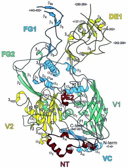
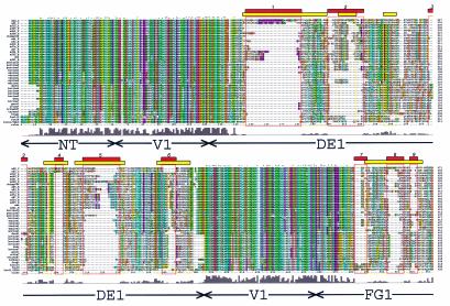
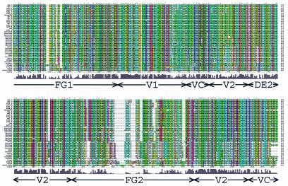
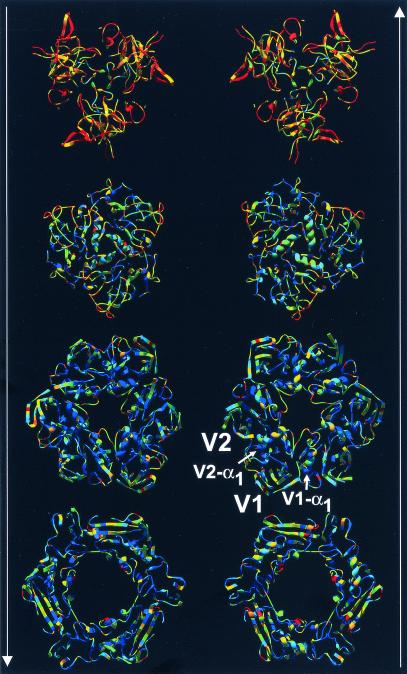
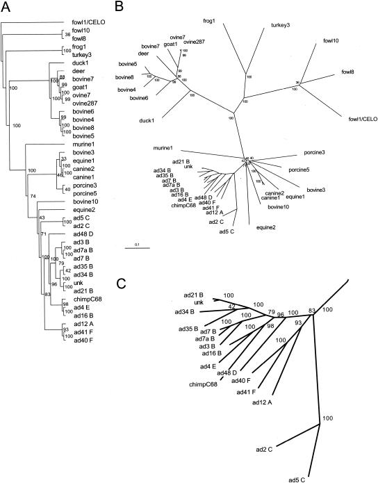
A major impediment to the use of adenovirus as a gene therapy vector and for vaccine applications is the host immune response to adenovirus hexon-the major protein component of the icosahedral capsid. A solution may lie in novel vectors with modified or chimeric hexons designed to evade the immune response. To facilitate this approach, we have distinguished the portion of hexon that all serotypes have in common from the hypervariable regions that are responsible for capsid diversity and type-specific immunogenicity. The common hexon core-conserved because it forms the viral capsid-sets boundaries to the regions where modifications can be made to produce nonnative hexons. The core has been defined from the large and diverse set of known hexon sequences by an accurate alignment based on the newly refined crystal structures of human adenovirus types 2 (Ad2) and Ad5 hexon. Comparison of the two hexon models, which are the most accurate so far, reveals that over 90% of the residues in each have three-dimensional positions that closely match. Structures for more distant hexons were predicted by building molecular models of human Ad4, chimpanzee adenovirus (AdC68), and fowl adenovirus 1 (FAV1 or CELO). The five structures were then used to guide the alignment of the 40 full-length (>900 residues) hexon sequences in public databases. Distance- and parsimony-based phylogenetic trees are consistent and reveal evolutionary relationships between adenovirus types that parallel those of their animal hosts. The combination of crystallography, molecular modeling, and phylogenetic analysis defines a conserved molecular core that can serve as the armature for the directed design of novel hexons.


Figures

FIG. 1.
FIG. 1.
Hexon structure. Ribbon representation of the Ad2 hexon subunit. The view is perpendicular to the molecular threefold axis from the inside of the molecule. The top of the molecule, which contains the loops (DE1, FG1, and FG2), forms the outer surface of the viral capsid. The hexon base contains a small loop (DE2) and two eight-stranded “viral” jellyrolls (V1 and V2), which are separated by the connector, VC. The eight jellyroll β strands are labeled B to I. The N-terminal loop, NT, lies underneath the base. The secondary structural elements are labeled, as are the positions of chain segments that are not observed in the crystallographic model (<…>). The figure was made with MOLSCRIPT (40).
FIG. 2.
FIG. 2.
Hexon sequence alignment. Multiple-sequence alignment of 40 hexon sequences calculated with Clustal_X (64, 65). An alignment profile based on the structural alignment of two hexon crystal structures and three homology models was used to guide the multiple-sequence alignment. The default Clustal_X color scheme indicates significant features. All G (orange) and P (yellow) are colored. Frequent occurrences of a property at a sequence position are colored: hydrophobic, blue; hydrophobic tendency, light blue; basic, red; acidic, purple; hydrophilic, green; unconserved, white. Conserved positions are marked as complete (*), strong (:), or weak (.). A histogram indicates the conservation level, and the eight molecular regions are labeled. Previously assigned (15) HVRs (HVR1 to -9) are boxed in yellow, and the new assignments (HVR1 to -9) are boxed in red. A Clustal_X alignment file is available for download at http://bioinfo.wistar.upenn.edu/pub/RKB_hexon.aln.
FIG. 2.
FIG. 2.
Hexon sequence alignment. Multiple-sequence alignment of 40 hexon sequences calculated with Clustal_X (64, 65). An alignment profile based on the structural alignment of two hexon crystal structures and three homology models was used to guide the multiple-sequence alignment. The default Clustal_X color scheme indicates significant features. All G (orange) and P (yellow) are colored. Frequent occurrences of a property at a sequence position are colored: hydrophobic, blue; hydrophobic tendency, light blue; basic, red; acidic, purple; hydrophilic, green; unconserved, white. Conserved positions are marked as complete (*), strong (:), or weak (.). A histogram indicates the conservation level, and the eight molecular regions are labeled. Previously assigned (15) HVRs (HVR1 to -9) are boxed in yellow, and the new assignments (HVR1 to -9) are boxed in red. A Clustal_X alignment file is available for download at http://bioinfo.wistar.upenn.edu/pub/RKB_hexon.aln.
FIG. 3.
FIG. 3.
Hexon trimer colored by sequence diversity. Layers of the trimeric hexon molecule displayed as ribbons. The two columns show hexon slices viewed along the molecular threefold axis. The arrows indicate the direction of view from either the virion exterior top to bottom (left) or from the virion interior bottom to top (right). The structures are colored by sequence diversity from blue (conserved) to yellow (less conserved) to red (not conserved). The jellyrolls V1 and V2 and their α1 helices are labeled. Note that the size of the hexon's pseudohexameric base is dictated by structural elements, such as V1-α1 and V2-α1, which separate the jellyrolls. A PDB file of the hexon trimer with temperature factors (B factors) representing sequence diversity is available for download at http://bioinfo.wistar.upenn.edu/pub/RKB_hexon_var.pdb.
FIG. 4.
FIG. 4.
Phylogenetic analysis of adenovirus hexon. Phylogenetic trees of 40 full-length hexon sequences calculated with the Phylip program package (23). (A) An unrooted tree generated by parsimony analysis with PROTPARS and CONSENSE. (B) An unrooted tree generated by distance matrix analysis with PROTDIST (Dayhoff's PAM 001 scoring matrix), FITCH (global rearrangements option), and CONSENSE. Branch lengths are proportional to the number of substitutions, and the scale bar represents 10 mutations per 100 sequence positions. Bootstrap values for the 100 trials are indicated for each branch. (C) A close-up view of the Hominidae adenovirus hexon branches.

Similar articles

Cited by

References

    1. Altschul, S. F., T. L. Madden, A. A. Schäffer, J. Zhang, Z. Zhang, W. Miller, and D. J. Lipman. 1997. Gapped BLAST and PSI-BLAST: a new generation of protein database search programs. Nucleic Acids Res. 25:3389-3402. - PMC - PubMed
    1. Arnberg, N., K. Edlund, A. H. Kidd, and G. Wadell. 2000. Adenovirus type 37 uses sialic acid as a cellular receptor. J. Virol. 74:42-48. - PMC - PubMed
    1. Athappilly, F. K., R. Murali, J. J. Rux, Z. Cai, and R. M. Burnett. 1994. The refined crystal structure of hexon, the major coat protein of adenovirus type 2, at 2.9 Å resolution. J. Mol. Biol. 242:430-455. - PubMed
    1. Bamford, D. H., R. M. Burnett, and D. I. Stuart. 2002. Evolution of viral structure. Theor. Pop. Biol. 61:461-470. - PubMed
    1. Benkö, M., B. Harrach, and J. C. D'Halluin. 1990. Molecular cloning and physical mapping of the DNA of bovine adenovirus serotype 4; study of the DNA homology among bovine, human, and porcine adenoviruses. J. Gen. Virol. 71:465-469. - PubMed

Publication types

LinkOut - more resources