. 2003 Sep;77(17):9567-77.
doi: 10.1128/jvi.77.17.9567-9577.2003.

Phylogenetic and evolutionary relationships among torovirus field variants: evidence for multiple intertypic recombination events

Affiliations

Phylogenetic and evolutionary relationships among torovirus field variants: evidence for multiple intertypic recombination events

S L Smits et al. J Virol. 2003 Sep.

Abstract

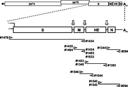
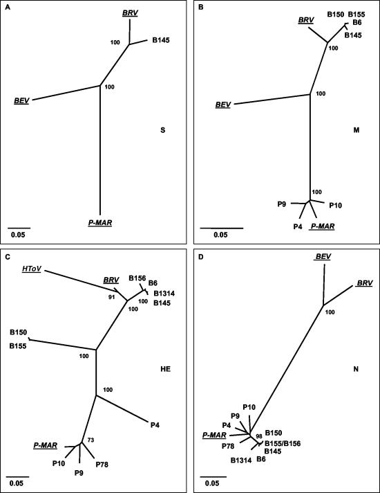
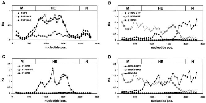
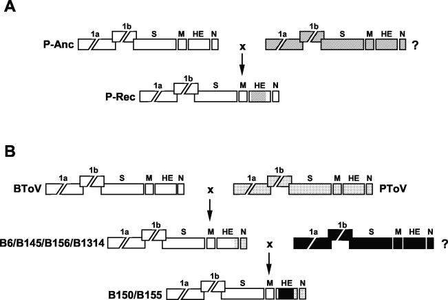
Toroviruses (family Coronaviridae, order Nidovirales) are enveloped, positive-stranded RNA viruses that have been implicated in enteric disease in cattle and possibly in humans. Despite their potential veterinary and clinical relevance, little is known about torovirus epidemiology and molecular genetics. Here, we present the first study into the diversity among toroviruses currently present in European swine and cattle herds. Comparative sequence analysis was performed focusing on the genes for the structural proteins S, M, HE, and N, with fecal specimens serving as sources of viral RNA. Sequence data published for animal and human torovirus variants were included. Four genotypes, displaying 30 to 40% divergence, were readily distinguished, exemplified by bovine torovirus (BToV) Breda, porcine torovirus (PToV) Markelo, equine torovirus Berne, and the putative human torovirus. The ungulate toroviruses apparently display host species preference. In phylogenetic analyses, all PToV variants clustered, while the recent European BToVs mostly resembled the New World BToV variant Breda, identified 19 years ago. However, we found ample evidence for recurring intertypic recombination. All newly characterized BToV variants seem to have arisen from a genetic exchange, during which the 3' end of the HE gene, the N gene, and the 3' nontranslated region of a Breda virus-like parent had been swapped for those of PToV. Moreover, some PToV and BToV variants carried chimeric HE genes, which apparently resulted from recombination events involving hitherto unknown toroviruses. From these observations, the existence of two additional torovirus genotypes can be inferred. Toroviruses may be even more promiscuous than their closest relatives, the coronaviruses and arteriviruses.


Figures

FIG. 1.
FIG. 1.
Electron microscopic detection of toroviruses in fecal specimens from diarrheic swine (P4 and P9) and cattle (B6). Virions were analyzed directly (B6) or after immunoaggregation with convalescent-phase serum from PToV P4-infected animals (P4 and P9). Negative staining was performed with 2% sodium phosphotungstic acid. Bars, 100 nm.
FIG. 2.
FIG. 2.
Torovirus genome organization and schematic outline of the strategies used for RT-PCR amplification of the genes for the structural proteins. The upper panel shows a schematic representation of the genome. Boxes represent the genes for the polymerase (ORF1a and ORF1b), the spike protein (S), the membrane protein (M), the hemagglutinin-esterase (HE), and the nucleocapsid protein (N). Open arrows indicate intergenic regions and transcription-regulating sequences. Also indicated are the cap structure (black dot) and the poly(A) tail (An). The lower panel shows a schematic outline of the RT-PCR assays employed to amplify S, M, HE, and N sequences. The orientations and positions of the oligonucleotides on the torovirus genome are shown.
FIG. 3.
FIG. 3.
Unrooted neighbor-joining trees depicting the phylogenetic relationships among torovirus field variants. Trees were constructed for (A) the S gene, (B) the M gene, (C) the HE gene, and (D) the N gene with the Kimura-2 parameter method. Confidence values calculated by bootstrap analysis (1,000 replicates) are indicated at the major branching points. Branch lengths are drawn to scale; the scale bar represents 0.05 nucleotide substitution per site. The torovirus reference strains BRV, BEV, P-MAR, and HToV are italicized and underlined. The tree shown for the HE gene was based upon an alignment corresponding to residues 1 to 1049 of B145. Note that BEV was not included in this tree; as a result of a large deletion, Berne virus has lost most of its HE gene (51).
FIG. 4.
FIG. 4.
Identification of recombination sites in the M, HE, and N genes of torovirus field variants. (A) Recombination sites were identified with the program PhylPro (58). This phylogenetic profile method introduces the phylogenetic correlation measure, i.e., the principle that phylogenetic relationships in different regions of an aligned sequence will be similar when no recombination has occurred. An alignment of the coding regions of the M, HE, and N genes of P-MAR, P4, P9, P10, BRV, B6, B145, B150, and B155 was generated. For each individual sequence in the alignment, the phylogenetic correlations were computed at every position with a sliding-window technique, with window limits fixed at 15 differences. Shown are the phylogenetic profiles of B6 and B145, with x and y axes indicating the nucleotide positions and the phylogenetic correlation, respectively; as a reference, the genes for M, HE, and N, drawn to scale, are depicted as boxes (top). For clarity, profiles of P4, B150, and B155 were hidden in the graph. (B) Phylogenetic profiles of B150 and B155; for clarity, profiles of P4, B6, and B145 were hidden. Window limits were fixed at 10 differences. (C) Alignments of HE sequences surrounding the recombination sites; arrows and numbers correspond to those in panels A and B. Nucleotide differences with respect to the B145 sequences are shown. Nucleotide positions given are numbered from A1 of the B145 HE gene.
FIG. 5.
FIG. 5.
Distribution of synonymous substitutions (Dss) in the M, HE, and N genes of torovirus field variants. Multiple alignments were generated for the protein-coding nucleotide sequences; all gaps were excluded. The sequences were compared pairwise by sliding-window analysis, during which the number of synonymous substitutions per synonymous site (Ks) was estimated for overlapping 240-nucleotide gene segments with a 60-nucleotide step size. Dss profiles were generated by plotting the calculated Ks values against nucleotide positions. As a reference, the genes for M, HE, and N, drawn to scale, are depicted as boxes (top). The Dss profiles shown were produced by pairwise comparison (A) of P4, P9, and P-MAR, (B) of B145 to BRV, P-MAR, or B6, (C) of B145 to B150 or B6 and of B150 to B6, and (D) of B150 to BRV or P-MAR, with the B145-B6 graph providing a baseline.
FIG. 6.
FIG. 6.
Hypothetical model for genetic exchanges among torovirus field variants. Torovirus genomes are depicted schematically, with the various genes represented by boxes. (A) Presumptive recombination event during PToV divergence, involving an ancestral PToV strain (P-Anc; genes indicated by white boxes) and an unknown toroviral parent (?; genes indicated by hatched boxes). Exchange of HE sequences resulted in recombinant progeny (P-Rec). (B) Presumptive recombination events during BToV divergence. During an initial single recombination event, the 3′ end of the HE gene and the N gene and the 3′-NTR of a BRV-like BToV variant (indicated in white) were replaced by the corresponding PToV sequences (dotted), giving rise to the B6/B145/B156/B1314 lineage. A subsequent double recombination event, during which BToV HE sequences were replaced by those of an as yet unidentified torovirus (?, indicated in black), resulted in the B150/B155 lineage.

Similar articles

Cited by

References

    1. Beards, G. M., D. W. Brown, J. Green, and T. H. Flewett. 1986. Preliminary characterisation of torovirus-like particles of humans: comparison with Berne virus of horses and Breda virus of calves. J. Med. Virol. 20:67-78. - PMC - PubMed
    1. Beards, G. M., C. Hall, J. Green, T. H. Flewett, F. Lamouliatte, and P. Du Pasquier. 1984. An enveloped virus in stools of children and adults with gastroenteritis that resembles the Breda virus of calves. Lancet i:1050-1052. - PMC - PubMed
    1. Boom, R., C. J. Sol, M. M. Salimans, C. L. Jansen, P. M. Wertheim-van Dillen, and J. van der Noordaa. 1990. Rapid and simple method for purification of nucleic acids. J. Clin. Microbiol. 28:495-503. - PMC - PubMed
    1. Brown, D. W., G. M. Beards, and T. H. Flewett. 1987. Detection of Breda virus antigen and antibody in humans and animals by enzyme immunoassay. J. Clin. Microbiol. 25:637-640. - PMC - PubMed
    1. Brown, D. W., R. Selvakumar, D. J. Daniel, and V. I. Mathan. 1988. Prevalence of neutralising antibodies to Berne virus in animals and humans in Vellore, South India. Arch. Virol. 98:267-269. - PMC - PubMed

MeSH terms

LinkOut - more resources