. 2003 Sep;77(17):9613-21.
doi: 10.1128/jvi.77.17.9613-9621.2003.

Point mutations in exon I of the herpes simplex virus putative terminase subunit, UL15, indicate that the most conserved residues are essential for cleavage and packaging

Affiliations

Point mutations in exon I of the herpes simplex virus putative terminase subunit, UL15, indicate that the most conserved residues are essential for cleavage and packaging

Angela J Przech et al. J Virol. 2003 Sep.

Abstract

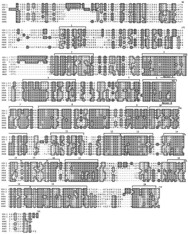
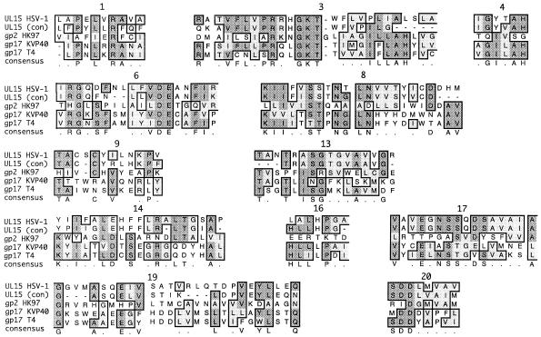
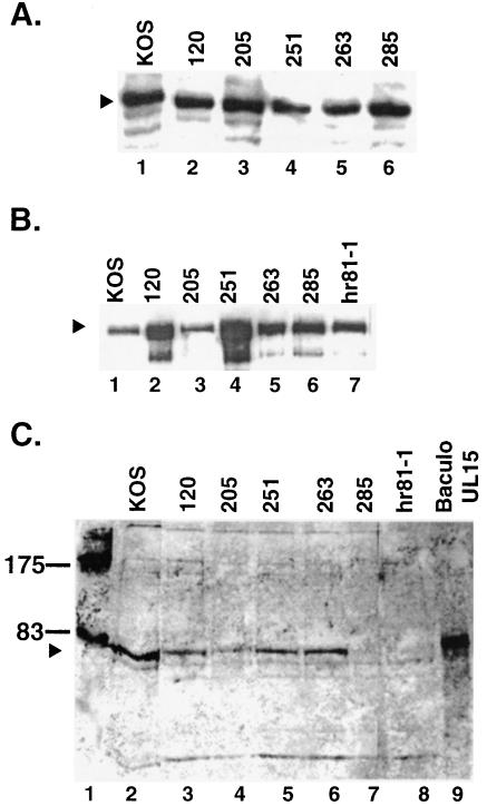
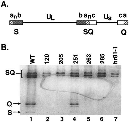
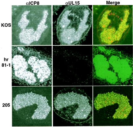
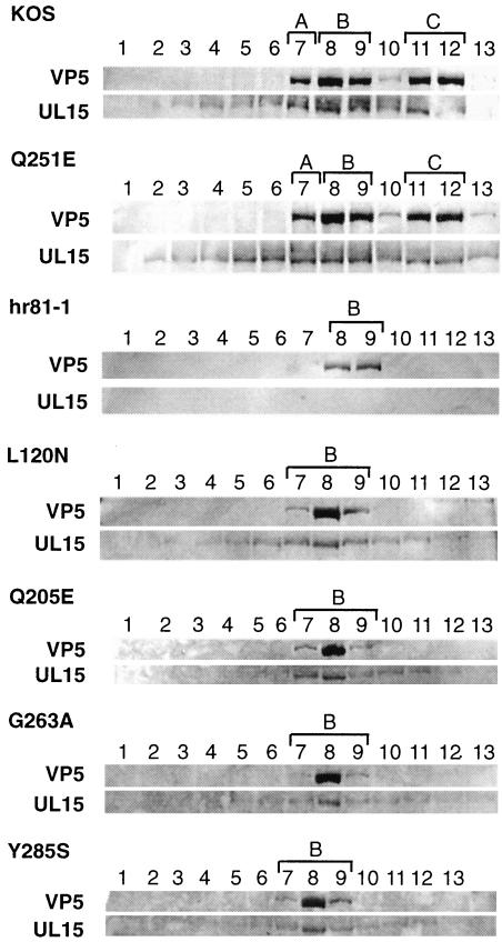
The herpes simplex virus UL15 and UL28 genes are believed to encode two subunits of the terminase involved in cleavage and packaging of viral genomes. Analysis of the UL15 protein sequence and its herpesvirus homologues revealed the presence of 20 conserved regions. Twelve of the twenty regions conserved among herpesviruses are also conserved in terminases from DNA bacteriophage. Point mutations in UL15 were designed in four conserved regions: L120N (CR1), Q205E (CR2), Q251E (CR3), G263A (CR3), and Y285S (CR4). Transfection experiments indicated that each mutant gene could produce stable UL15 protein at wild-type levels; however, only one mutant (Q251E) was able to complement the UL15-null virus. Each mutation was introduced into the viral genome by marker transfer, and all mutants except Q251E were unable to form plaques on Vero cells. Furthermore, failure to form plaques on Vero cells correlated with a defect in cleavage and packaging. Immunofluorescence experiments indicated that in cells infected with all mutant viruses the UL15 protein could be detected and was found to localize to replication compartments. Although wild-type and mutant Q251E were able to produce A, B, and C capsids, the rest of the mutants were only able to produce B capsids, a finding consistent with their defects in cleavage and packaging. In addition, all mutant UL15 proteins retained their ability to interact with B capsids. Therefore, amino acid residues 120, 205, 263, and 285 are essential for the cleavage and packaging process rather than for association with capsids or localization to replication compartments.


Figures

FIG. 1.
FIG. 1.
UL15 homology alignments: UL15 is highly conserved among the herpesvirus family. A PSI-BLAST search was performed on the HSV-1 UL15 protein sequence to obtain all its homologues. The eight human herpesvirus homologues were aligned using CLUSTALW (Blosum algorithm) within MacVector. We assigned 20 CRs between UL15 and its homologues based on the fact that many but not all of these regions are also conserved in bacteriophage terminase proteins (see Fig. 2). The Walker A and B boxes of the putative ATP-binding domain are indicated.
FIG. 2.
FIG. 2.
UL15 and phage alignments. A PSI-BLAST search was performed on gp2 from HK97. UL15 (HSV-1), the UL15 consensus sequence from Fig. 1 [UL15(con)], gp2 (HK97), gp17v (KVP40), and gp17 (T4) were aligned by using CLUSTALW (identity algorithm) within MacVector. Of the 20 CRs shown in Fig. 1, 12 are also conserved between HSV and the phage terminases. The numbers above each CR correspond to the CRs shown in Fig. 1.
FIG. 3.
FIG. 3.
UL15 detected by Western blot analysis. (A) Western blot analysis of transiently transfected cells. Vero cells (2.1 × 10⁶) transfected with a pcDNA plasmid containing a wild-type or mutant version of the UL15 gene were harvested 20 h posttransfection, subjected to SDS-PAGE on a 12% gel, and transferred to an ECL membrane. Western blot analysis was performed by using αUL15 (1:2,000) and AP Immunestar development (Bio-Rad). The arrowhead indicates the band corresponding to the 81-kDa UL15 protein (lane 1). (B and C) Western blot analysis of infected cell lysates. Vero cells (2.1 × 10⁶) were infected with wild-type or mutant virus at an MOI of 10 PFU/cell and harvested 20 h postinfection. The samples were subjected to SDS-PAGE on a 12% gel and then transferred to an ECL membrane. Western blot analysis was performed by using αVP5 (1:2,000) and ECL development (Amersham Pharmacia) (B) and αUL15 (1:2,000) and AP development (Promega) (C). In panel B, the arrowhead indicates the position of VP5. In panel C, the arrowhead indicates the position of the 81-kDa form of UL15. An extract from insect cells infected with a UL15 expressing baculovirus was used as a control for the presence of the 81-kDa form of UL15 (lane 9). The positions of molecular mass markers (in kilodaltons) are shown in lane 1.
FIG. 4.
FIG. 4.
Southern blot analysis of viral DNA from cells infected with wild-type and mutant viruses. (A) The HSV-1 genome consists of two unique regions, UL and US, which are flanked by repeated sequences a, b, and c. The subscript n in “an” indicates multiple copies of the a sequence. The BamHI fragments corresponding to S, Q, and SQ junction fragments are indicated. (B) Vero cells (107) were infected with the indicated virus at an MOI of 10 PFU/cell for 18 h. Total DNA was digested with BamHI, subjected to electrophoresis, and transferred to a GeneScreen Plus membrane. The membrane was probed with a 32P-labeled BamHI SQ junction fragment. Arrows indicate the junction fragments SQ, Q, and S (from top to bottom). DNA from cells infected with wild-type and null mutant virus (hr81-1) are shown in lanes 1 and 7, respectively. Lanes 2 to 6 contained DNA from cells infected with mutants L120N, Q205E, Q251E, G263A, and Y285S, respectively. Although mutants L120N, Q205E, and hr81-1 appear to contain a reduced amount of SQ DNA compared to wild type and the other mutants, this difference was not reproducible, and we do not feel that it reflects a true reduction in levels of viral DNA synthesis.
FIG. 5.
FIG. 5.
Immunofluorescence analysis. Vero cells were infected with wild-type virus (KOS) (top row), UL15-null virus (hr81-1) (middle row), or the UL15 mutant virus Q205E (bottom row) at an MOI of 10 PFU/cell and fixed for immunofluorescence at 8 h postinfection. Infected cells were stained with anti-ICP8 monoclonal antibody (39S) (left panels, green) and anti-UL15 polyclonal antibody (αAS9) (middle panels, red). The right-hand column labeled Merge shows the merged images. The color was adjusted on the hr81-1 panels to demonstrate the lack of UL15 in these cells.
FIG. 6.
FIG. 6.
Western blot analysis of wild-type and mutant capsids. MV cells were infected with the indicated virus at an MOI of 5 PFU/cell for 20 h. Sucrose gradient fractions were collected as described in Materials and Methods. Gradient fractions for each virus were subjected to SDS-PAGE on a 10% gel, and Western blot analysis was performed by using αAS9 (αUL15) and AP development (Promega) as described in Materials and Methods. The αAS9 antibody cross-reacts with VP5 proteins, and this cross-reaction aids in the detection of fractions that contain capsids (top panel for each virus). We previously reported an 87-kDa protein which associated with C capsids that was detected by αAS9 (29, 37). In the experimental conditions used in the present study, the 87-kDa protein was not observed. The bottom panel for each virus shows the position of the 81-kDa form of UL15. Fractions containing A, B, and C capsids are marked with brackets.

Similar articles

Cited by

References

    1. Abbotts, A. P., V. G. Preston, M. Hughes, A. H. Patel, and N. D. Stow. 2000. Interaction of the herpes simplex virus type 1 packaging protein UL15 with full-length and deleted forms of the UL28 protein. J. Gen. Virol. 81:2999-3009. - PubMed
    1. Adelman, K., B. Salmon, and J. D. Baines. 2001. Herpes simplex virus DNA packaging sequences adopt novel structures that are specifically recognized by a component of the cleavage and packaging machinery. Proc. Natl. Acad. Sci. USA 98:3086-3091. - PMC - PubMed
    1. al-Kobaisi, M. F., F. J. Rizon, I. McDougall, and V. G. Preston. 1991. The herpes simplex virus UL33 gene product is required for the assembly of full capsids. J. Virol. 180:380-388. - PubMed
    1. Altschul, S. F., T. L. Madden, A. A. Schaffer, J. Zhang, Z. Zhang, W. Miller, and D. J. Lipman. 1997. Gapped BLAST and PSI-BLAST: a new generation of protein database search programs. Nucleic Acids Res. 25:3389-3402. - PMC - PubMed
    1. Baines, J. D., A. P. Poon, J. Rovnak, and B. Roizman. 1994. The herpes simplex virus 1 UL15 gene encodes two proteins and is required for cleavage of genomic viral DNA. J. Virol. 68:8118-8124. - PMC - PubMed

Publication types

MeSH terms

LinkOut - more resources