. 2003 Sep;77(17):9685-94.
doi: 10.1128/jvi.77.17.9685-9694.2003.

A bromodomain-containing protein from tomato specifically binds potato spindle tuber viroid RNA in vitro and in vivo

Affiliations

A bromodomain-containing protein from tomato specifically binds potato spindle tuber viroid RNA in vitro and in vivo

Angel Emilio Martínez de Alba et al. J Virol. 2003 Sep.

Abstract

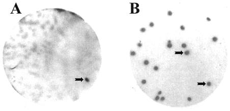
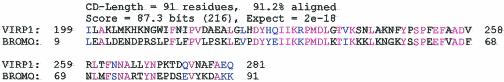
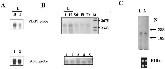
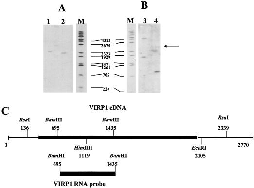
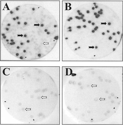
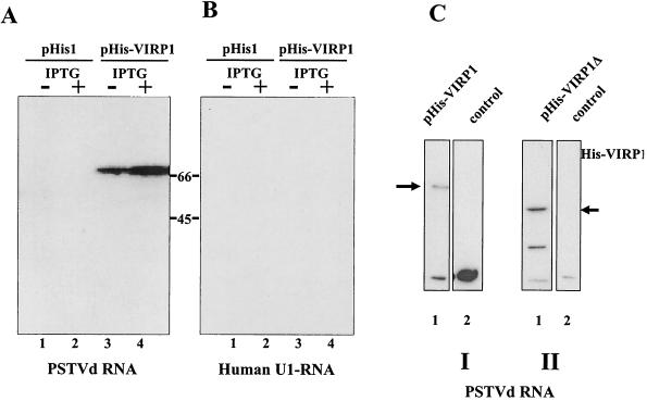
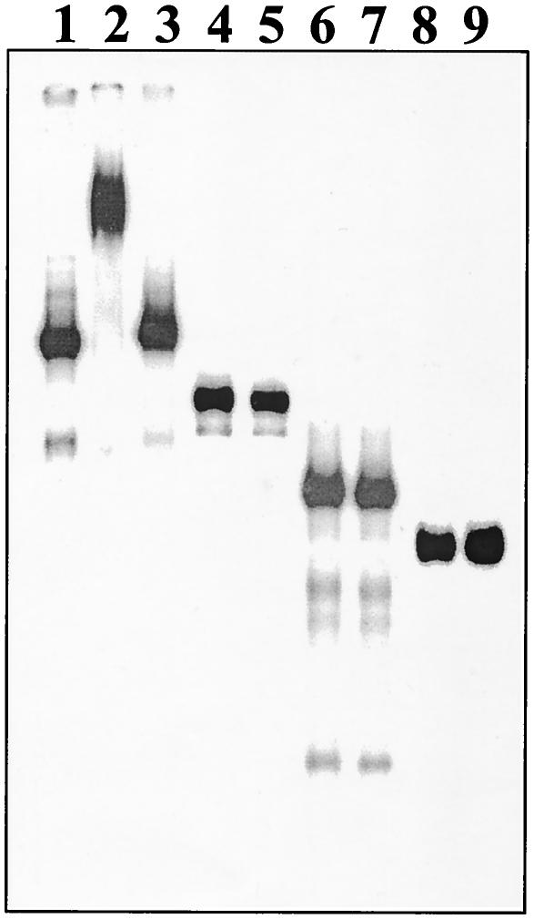
For the identification of RNA-binding proteins that specifically interact with potato spindle tuber viroid (PSTVd), we subjected a tomato cDNA expression library prepared from viroid-infected leaves to an RNA ligand screening procedure. We repeatedly identified cDNA clones that expressed a protein of 602 amino acids. The protein contains a bromodomain and was termed viroid RNA-binding protein 1 (VIRP1). The specificity of interaction of VIRP1 with viroid RNA was studied by different methodologies, which included Northwestern blotting, plaque lift, and electrophoretic mobility shift assays. VIRP1 interacted strongly and specifically with monomeric and oligomeric PSTVd positive-strand RNA transcripts. Other RNAs, for example, U1 RNA, did not bind to VIRP1. Further, we could immunoprecipitate complexes from infected tomato leaves that contained VIRP1 and viroid RNA in vivo. Analysis of the protein sequence revealed that VIRP1 is a member of a newly identified family of transcriptional regulators associated with chromatin remodeling. VIRP1 is the first member of this family of proteins, for which a specific RNA-binding activity is shown. A possible role of VIRP1 in viroid replication and in RNA mediated chromatin remodeling is discussed.


Figures

FIG. 1.
FIG. 1.
Detection of PSTVd RNA-binding protein VIRP1. A total of about 600,000 plaques from a cDNA expression library from tomato leaves were subjected to a primary screening, by using a longer-than-unit-length PSTVd RNA transcript (57) as a radioactively labeled probe. (A) The tomato cDNA expression library was plated at ca. 5,000 PFU/plate. The black arrow shows the signal corresponding to the viroid binding protein 1 clone after a primary screening. (B) The clone picked from the primary screening was plated at a density of 25 PFU/plate, and the PSTVd RNA-binding properties of positive clone 1 were confirmed by secondary screening. Plaque lift and binding assays with α-32P-labeled RNA transcripts were performed as described in Materials and Methods.
FIG. 2.
FIG. 2.
Bromodomain of VIRP1. Consensus protein motif pfam00439 (protein motif data collection Pfam 8.0) is aligned with a region of VIRP1. The data were retrieved from http://www.ncbi.nlm.nih.gov/Structure/cdd/wrpsb.cgi. Identical amino acids are in red, and the conservative changes are in blue.
FIG. 3.
FIG. 3.
Expression and tissue distribution of VIRP1 mRNA in tomato. mRNA extracted from 100 μg of total RNA was size separated in 1% agarose-formaldehyde (A and C) or agarose-guanidine thiocyanate (B) gels. RNA was blotted and hybridized with an α-32P-labeled 0.7-kb VIRP1 antisense RNA probe. (A) Northern blot of RNAs from healthy (lane H) and PSTVd-infected (lane I) tomato leaves. The same blot was also probed with an actin RNA transcript for loading controls as shown in the lower panel (lanes 1 and 2, respectively). (B) Northern blots of RNAs from various tissues. Poly(A)+ mRNA from leaf (L, lanes I [infected] and H [healthy]), seed (lane Sd), flower (lane Fl), and fruit (Fr) were hybridized with antisense VIRP1 RNA. M, DNA molecular weight marker. (C) Northern blot of total RNA from petals (lane 1) and sepals (lane 2) of infected plants hybridized with VIRP1 DNA. Equal loading was verified by visualization of 28S and 18S rRNAs on the blot.
FIG. 4.
FIG. 4.
Southern analysis of tomato genomic DNA. (A and B) Southern analysis of tomato total DNA. Lane 1, digestion with RsaI; lane 2, digestion with RsaI/EcoRI; lane 3, digestion with HindIII; lane 4, digestion with BamHI; lane M, lambda DNA molecular weight marker digested with BstEII. (C) Restriction map of VIRP1 and the probe used for hybridization. The coding region is shown by a heavy black box.
FIG. 5.
FIG. 5.
Binding of VIRP1 by plaque lift assay. Binding specificity of VIRP1 and PSTVd RNA was tested by plaque lift assay. A mixture (1:1) of λVIRP1 and λ-ZAPII phage was plated out and tested for binding with different RNAs. (A and B) Only when monomeric (A) or multimeric (B) PSTVd positive-strand RNA forms (for details see Materials and Methods) were used as RNA probes could positive signals be discriminated from those due to background. (C and D) When the same mixture was plated at a lower density of 25 PFU/plate and allowed to interact with either potato U1 RNA (C) or human U1 RNA (D), only background signals were visible. In panels C and D the numbers of signals visible on the autoradiograph were the same as the numbers of plaques per plate. The black arrows show the signals corresponding to VIRP1 plaques, which interact with PSTVd (positive signals), while the open arrows indicate the background signals.
FIG. 6.
FIG. 6.
Northwestern analysis of VIRP1. Specificity of binding of VIRP1 protein with PSTVd positive-strand RNA was confirmed by Northwestern assay. Crude extracts of E. coli BL21 cells harboring either pHIS, pHis-VIRP1 or pHis1-VIRP1Δ or without plasmid (see the text for details) were separated by SDS-10% PAGE, transferred to nitrocellulose membranes and probed with radioactively labeled transcripts. In panels A and C, hybridization was done with PSTVd positive-strand RNA; in panel B, hybridization was done with U1 RNA. (A and B) BL21(pHIS) or BL21(pHIS-VIRP1) crude cell extracts. Lanes 1 and 3, noninduced cells; lanes 2 and 4, IPTG-induced cells. (C) BL21 cell extracts. Lane I1, BL21(pHis-VIRP1) cell extracts; lane I2, BL21 cell extracts; lane II1, BL21(pHis1-VIRP1Δ) cell extracts; lane II2, BL21 cell extracts. The arrows indicate the VIRP1 and VIRP1Δ proteins, respectively. In panel C, parts I and II present results from two different experiments.
FIG. 7.
FIG. 7.
Specificity of binding of PSTVd RNA by EMSA. Radioactively labeled RNAs were incubated with or without purified VIRP1Δ protein for 60 min at room temperature. Mixtures were electrophoresed on 6% native polyacrylamide gels and subjected to autoradiography. When PSTVd RNA (lanes 1 to 3) was incubated with VIRP1Δ protein (lane 2), retardation due to RNA-protein complex formation could be observed, which could be competed for in the presence of 100-fold excess nonlabeled PSTVd RNA (lane 3). In contrast, when other RNA transcripts, e.g., pGEM-3Zf(−) (lanes 4 and 5), potato U1 snRNA (lanes 6 and 7), or pBluescript II KS(+) (lanes 8 and 9), were incubated together with VIRP1Δ (lanes 5, 7, and 9), no such retardation was detected.
FIG. 8.
FIG. 8.
VIRP1-PSTVd RNA in vivo complexes. PSTVd-infected extracts from irradiated tissue were used for IP assays. After IP, the samples were treated with proteinase K, and RNAs were recovered by ethanol precipitation after phenolization. Lane 1, RNAs extracted from IP reaction with a VIRP1Δ-specific polyclonal antibody; lane 2, nonspecific polyclonal antibody used for the IP; lane 3, no antibody was used. Extracted RNAs were separated by PAGE, electroblotted, and hybridized with a α-32P-labeled negative-strand PSTVd RNA probe.

Similar articles

Cited by

References

    1. Barlev, N. A., V. Poltoratsky, T. Owen-Hughes, C. Ying, L. Liu, J. L. Workman, and S. L. Berger. 1998. Repression of GCN5 histone acetyltransferase activity via bromodomain-mediated binding and phosphorylation by the Ku-DNA-dependent protein kinase complex. Mol. Cell. Biol. 18:1349-1358. - PMC - PubMed
    1. Beck, S., I. Hanson, A. Kelly, D. J. Pappin, and J. Trowsdale. 1992. A homologue of the Drosophila female sterile homeotic (fsh) gene in the class II region of the human MHC. DNA Seq. 2:203-210. - PubMed
    1. Branch, A. D., B. J. Benenfeld, and H. D. Robertson. 1988. Evidence for a single rolling circle in the replication of potato spindle tuber viroid. Proc. Natl. Acad. Sci. USA 85:9128-9132. - PMC - PubMed
    1. Chamberlin, H. M., and J. H. Thomas. 2000. The bromodomain protein LIN-49 and trithorax-related protein LIN-59 affect development and gene expression in Caenorhabditis elegans. Development 127:713-723. - PubMed
    1. Daros, J. A., and R. Flores. 2002. A chloroplast protein binds a viroid RNA in vivo and facilitates its hammerhead-mediated self-cleavage. EMBO J. 21:749-759. - PMC - PubMed

Publication types

MeSH terms

Associated data