. 2003 Sep 2;100(18):10411-6.
doi: 10.1073/pnas.1834278100. Epub 2003 Aug 12.

Rapamycin causes down-regulation of CCR5 and accumulation of anti-HIV beta-chemokines: an approach to suppress R5 strains of HIV-1

Affiliations

Rapamycin causes down-regulation of CCR5 and accumulation of anti-HIV beta-chemokines: an approach to suppress R5 strains of HIV-1

A Heredia et al. Proc Natl Acad Sci U S A. .

Abstract

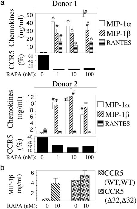
Propagation of R5 strains of HIV-1 on CD4 lymphocytes and macrophages requires expression of the CCR5 coreceptor on the cell surface. Individuals lacking CCR5 (CCR5 Delta 32 homozygous genotype) are phenotypically normal and resistant to infection with HIV-1. CCR5 expression on lymphocytes depends on signaling through the IL-2 receptor. By FACS analysis we demonstrate that rapamycin (RAPA), a drug that disrupts IL-2 receptor signaling, reduces CCR5 surface expression on T cells at concentrations as low as 1 nM. In addition, lower concentrations of RAPA (0.01 nM) were sufficient to reduce CCR5 surface expression on maturing monocytes. PCR analysis on peripheral blood mononuclear cells (PBMCs) showed that RAPA interfered with CCR5 expression at the transcriptional level. Reduced expression of CCR5 on PBMCs cultured in the presence of RAPA was associated with increased extracellular levels of macrophage inflammatory protein (MIP)-1 alpha and MIP-1 beta. In infectivity assays, RAPA suppressed the replication of R5 strains of HIV-1 both in PBMC and macrophage cultures. In total PBMC cultures, RAPA-mediated inhibition of CCR5-using strains of HIV-1 occurred at 0.01 nM, a concentration of drug that is approximately 103 times lower than therapeutic through levels of drug in renal transplant recipients. In addition, RAPA enhanced the antiviral activity of the CCR5 antagonist TAK-779. These results suggest that low concentrations of RAPA may have a role in both the treatment and prevention of HIV-1 infection.


Figures

Fig. 1.
Fig. 1.
Effect of RAPA on proliferation of PBMCs. Purified PBMCs from normal donors were cultured in the presence of IL-2 and RAPA. On day 7, the extent of cell proliferation was measured by the MTT assay. Representative results obtained on one of two independent experiments, each using cells from four donors, are shown. For each donor, data values are mean ± SD of three independent wells.
Fig. 2.
Fig. 2.
RAPA down-regulates CCR5 expression on T cells and monocytes. (a) Staining experiment showing specific detection of CCR5 surface expression on CD4+ T cells from a normal donor, but not on CD4+ T cells from an individual homozygous for the Δ32 mutation in the CCR5 gene. (b) Down-regulation of CCR5 surface expression on CD4+ T cells by RAPA. PBMCs cultured for 7 days in the presence of IL-2 and RAPA were assayed for CCR5 levels. Expression of CCR5 on CD4+ T lymphocytes is shown as a solid line, and fluorescence due to the IgG isotype control is shown as a dashed line. (c) Inhibition of CCR5 mRNA transcription in PBMCs by RAPA. Total RNA was isolated from PBMCs that had been cultured in the presence of IL-2 and RAPA for 7 days (cells from same experiment as on b). Equivalent amounts of RNA were subjected to RT-PCR using primer pairs specific for the amplification of CCR5 mRNA (Upper) and 18S ribosomal RNA (Lower). (d) RAPA down-regulates CCR5 cell-surface expression on maturing monocytes. Monocytes cultured for 5 days in the presence of RPMI 20/10% ABHS and RAPA were dually immunostained for CD14 and CCR5. Changes in CCR5 surface expression were examined in CD14-gated cells. The immunofluorescence profile obtained with the anti-CCR5 mAb 182 (solid line) is compared with that of the IgG2b isotype control (dashed line). Results in b and c are representative of data obtained in PBMCs from five different donors. Results in d are representative of similar profiles obtained on three different donors.
Fig. 3.
Fig. 3.
RAPA increases extracellular β-chemokine levels in PBMC cultures. (a) Donor PBMCs were cultured in the presence of IL-2 and RAPA for 10 days, at which time supernatants were evaluated for β-chemokine content by ELISA and cells were stained for CCR5 expression. Percentage of CD4 lymphocytes expressing CCR5 at each concentration of RAPA is indicated. Results shown in two donors are representative of four experiments using four different donors. *, P < 0.01; #, P < 0.05, compared with untreated control by Student's t test. (b) Effect of RAPA on extracellular levels of MIP-1β in cultures of CCR5-null PBMCs. Levels of MIP-1β protein in the presence and absence of RAPA were measured in supernatants of IL-2-stimulated PBMCs from a normal donor and from a donor homozygous for the Δ32 mutation in the CCR5 gene. Values were obtained on day 10 of culture and are means ± SD of duplicate wells. Results are representative of two independent experiments, each using cells from two normal donors.
Fig. 4.
Fig. 4.
RAPA inhibits HIV-1 replication in PBMCs, and the antiviral activity in R5 HIV-1 is greater than in X4 HIV-1. (a) Seven-day RAPA-treated PBMCs were infected with HIV-1 IIIb or HIV-1 ADA. Infected cells were cultured in the presence of drug for 7 days, at which time virus replication was measured by p24 and cell viability was measured by the MTT assay. Results (means ± SD of triplicate wells) are representative of seven independent experiments, each on cells from a different donor. (b) DNase-treated stocks of HIV-1 IIIb and HIV-1 ADA were used to infect PBMCs that had been treated with or without 100 nM RAPA. HIV-1 DNA sequences were amplified by PCR in cellular lysates prepared 24 h after infection. Amplified PCR products were detected with a radioactive probe. + indicates presence of RAPA in the PBMC culture before and after infection; - indicates no RAPA treatment. Amplification of β-actin sequences indicated same amount of cellular DNA among the different cell lysates (data not shown). NC denotes PCR negative control. (c) The antiviral activity of low concentrations of RAPA was investigated in a panel of R5 strains of HIV-1. Cell proliferation was assayed on uninfected cells from same donor cultured under identical conditions. Results (means ± SD of triplicate wells) are representative of three independent experiments, each on different donor cells.
Fig. 5.
Fig. 5.
RAPA inhibits HIV-1 replication in MDMs. Purified monocytes were cultured for 5 days in the presence of RAPA. On day 5, cells were infected with HIV-1 ADA and cultured in the presence of RAPA for 14 additional days. On days 7, 10, and 14 after infection, virus growth was measured by the RT assay. On day 14, cell viability was determined by MTT. Results (means ± SD) are representative of data obtained in three independent experiments, each using cells from a different donor.
Fig. 6.
Fig. 6.
RAPA enhances the antiviral activity of the CCR5 antagonist TAK-779. PBMCs that had been cultured in the absence or presence of RAPA (1, 10, and 100 nM) for 7 days were infected with HIV-1 ADA in the presence of 0.1 nM TAK-779. Infected cells were cultured in the presence of RAPA and 0.1 nM TAK-779. On day 7 after infection, virus production was measured by the p24 assay in the culture supernatant. Note the logarithmic scale in the y axis. Data represent means ± SD of triplicate wells. Representative results obtained in one of three independent experiments are shown.

Similar articles

Cited by

References

    1. Liu, R., Paxton, W., Choe, S., Ceradini, D., Martin, S., Horuk, R., MacDonald, M., Stuhlmann, H., Koup, R. & Landau, N. (1996) Cell 86, 367-377. - PubMed
    1. Lin, Y.-L., Mettling, C., Portales, P., Reynes, J., Clot, J. & Corbeau, P. (2002) Proc. Natl. Acad. Sci. USA 99, 15590-15595. - PMC - PubMed
    1. Cocchi, F., DeVico, A., Garzino-Demo, A., Arya, S., Gallo, R. & Lusso, P. (1995) Science 270, 1811-1815. - PubMed
    1. Baba, M., Nishimura, O., Kanzaki, N., Okamoto, M., Sawada, H., Iizawa, Y., Shiraishi, M., Aramaki, Y., Okonogi, K., Ogawa, Y., et al. (1999) Proc. Natl. Acad. Sci. USA 96, 5698-5703. - PMC - PubMed
    1. Strizki, J., Xu, S., Wagner, N., Wojcik, L., Liu, J., Hou, Y., Endres, M., Palani, A., Shapiro, S., Clader, J. W., et al. (2001) Proc. Natl. Acad. Sci. USA 98, 12718-12723. - PMC - PubMed