

Detection of human papillomavirus-16 in ovarian malignancy
- PMID: 12915876
- PMCID: PMC2376933
- DOI: 10.1038/sj.bjc.6601172
Detection of human papillomavirus-16 in ovarian malignancy
Abstract
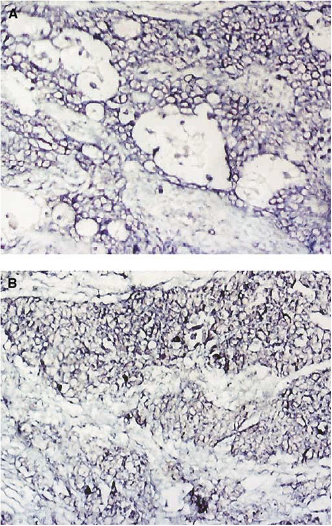
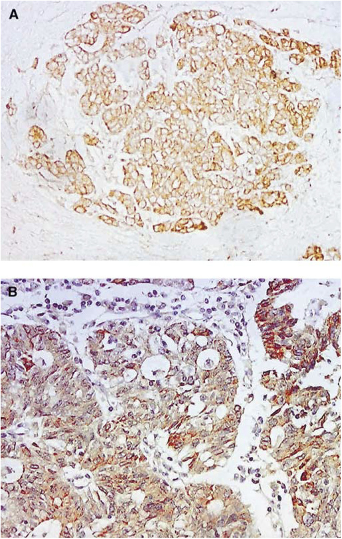
Human papillomavirus is the causal factor for cervical cancer. However, the role of HPV infection in ovarian cancer is unclear. This study aimed to determine the presence of human papillomavirus-16 (HPV-16) in ovarian cancer tissues. Archived human ovarian cancer tissues (N=54 cases, 50 are epithelial cancer, four are nonepithelial cancer) embedded in paraffin blocks were used. Controls are 30 nonmalignant ovarian tissue blocks. In situ hybridisation (ISH) and immunohistochemistry (IHC) were used to detect the presence of HPV-16 and p53 expression. In all, 52 or 36% of the epithelial ovarian tumours detected by ISH or IHC, respectively, were HPV-16 E6 positive. In contrast, only 6.7% of normal ovarian tissues were HPV-16 positive proved by ISH. Human papillomavirus-16 infection was significantly higher in cancer tissues compared to controls with an odds ratio of 16.7 (95% confidence interval [CI]=3.2-71.4, P<0.01). No significant correlation between HPV-16 infection and histological types of cancer was found (P>0.05). p53 gene expression was detected in 42% epithelial ovarian cancers. No correlation between p53 expression and HPV-16 infection was found. The results showed the presence of HPV-16 E6 in ovarian carcinoma, suggesting that HPV infection might play a role in ovarian carcinogenesis.
Figures
References
-
- Anttila M, Syrjanen S, Ji H, Saarikoski S, Syrjanen K (1999) Failure to demonstrate human papillomavirus DNA in epithelial ovarian cancer by general primer PCR. Gynecol Oncol 72: 337–341 - PubMed
-
- Anwar K, Nakakuki K, Imai H, Shiraishi T, Inuzuka M (1996) Infection of human papillomavirus (HPV) and p53 over-expression in human female genital tract carcinoma. J Pak Med Assoc 46: 220–224 - PubMed
-
- Beckmann AM, Sherman KJ, Saran L, Weiss NS (1991) Genital-type human papillomavirus infection is not associated with surface epithelial ovarian carcinoma. Gynecol Oncol 43: 247–251 - PubMed
-
- Boyle P, Maisonneuve P, Autier P (2000) Update on cancer control in women. Int J Gynecol Obstet 70: 263–303 - PubMed
MeSH terms
Substances
LinkOut - more resources
Full Text Sources
Other Literature Sources
Medical
Research Materials
Miscellaneous
