

Case Reports
Imaging characteristics of diffuse primary cutaneous B-cell lymphoma of the cranial vault with orbital and brain invasion
Affiliations
- PMID: 12917120
- PMCID: PMC7973699
Item in Clipboard
Case Reports
Imaging characteristics of diffuse primary cutaneous B-cell lymphoma of the cranial vault with orbital and brain invasion
AJNR Am J Neuroradiol.
2003 Aug.
Abstract
We herein present the imaging findings in a case of diffuse primary cutaneous B-cell lymphoma of the cranial vault with orbital and brain invasion. MR imaging revealed first concomitant orbital and parenchymal invasion in a case of primary diffuse non-Hodgkin lymphoma of the cranial vault. Contrast-enhanced MR imaging revealed a diffuse mass in the scalp of the frontoparietal region bilaterally and invasion of the right orbit and left frontoparietal lobe. Histologic and immunopathologic examinations revealed diffuse primary cutaneous B-cell lymphoma.
Figures
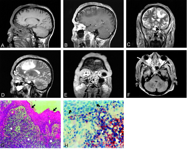
Images of a 65-year-old man who presented with a 6-month history of a right frontoparietal nodule that included ulcers and a 1-month history of swelling on the right lid. A, Unenhanced sagittal T1-weighted MR image does not show any cranial invasion. B, Contrast-enhanced sagittal T1-weighted MR image shows strong enhancement of a diffuse mass in the scalp bilateral frontoparietal region and invasion of the dura and left gyrus frontalis medius. C, Coronal view T2-weighted image shows isointense lesion and peripheral hyperintense edema on the left medial frontal gyrus. D, Sagittal T2-weighted image shows hyperintense edema on the frontal lobe. E, Contrast-enhanced coronal T1-weighted image shows marked enhancement at the right lateral orbital wall and muscle rectus lateralis. F, Axial fluid-attenuated inversion recovery image shows invasion of orbital wall (arrow) and muscle (arrowheads). G, Photomicrograph of a specimen shows tumoral cells (stars) in stratified squamous epithelium (arrows) (hematoxylin and eosin; original magnification, ×200). H, Photomicrograph shows CD20-positive tumoral cells (arrowheads) (strep ABC; original magnification, ×100) and stratified squamous epithelium (arrows).
Similar articles
-
Non-Hodgkin lymphoma of the cranial vault with retrobulbar metastasis mimicking a subacute subdural hematoma: case report.J Neurosurg. 2008 May;108(5):1018-20. doi: 10.3171/JNS/2008/108/5/1018. J Neurosurg. 2008. PMID: 18447722
-
Primary immunoblastic B-cell lymphoma of the cranial vault.Acta Neurochir (Wien). 2008 May;150(5):507-8; discussion 508. doi: 10.1007/s00701-007-1416-6. Epub 2008 Feb 25. Acta Neurochir (Wien). 2008. PMID: 18297234
-
Primary malignant non-Hodgkin's lymphoma of cranial vault: a case report.Surg Neurol. 1993 Apr;39(4):286-9. doi: 10.1016/0090-3019(93)90007-n. Surg Neurol. 1993. PMID: 8488447 Review.
-
Diffuse primary non-Hodgkin's lymphoma of the cranial vault.Br J Neurosurg. 2004 Oct;18(5):518-23. doi: 10.1080/02688690400012491. Br J Neurosurg. 2004. PMID: 15799158 Review.
-
Teaching NeuroImages: Primary diffuse large B-cell lymphoma of the cranial vault.Neurology. 2009 Oct 27;73(17):e84-5. doi: 10.1212/WNL.0b013e3181bd8283. Neurology. 2009. PMID: 19858452 No abstract available.
Cited by
-
A 60-year-old Indian male with altered sensorium and extensive lymphoma of the scalp.Semin Oncol. 2013 Jun;40(3):e9-21. doi: 10.1053/j.seminoncol.2013.04.014. Semin Oncol. 2013. PMID: 23806503 Free PMC article. No abstract available.
-
Characteristics of cranial vault lymphoma from a systematic review of the literature.Surg Neurol Int. 2022 Jun 3;13:231. doi: 10.25259/SNI_28_2022. eCollection 2022. Surg Neurol Int. 2022. PMID: 35855149 Free PMC article. Review.
-
Massive transcalvarial lymphoma.BMJ Case Rep. 2009;2009:bcr2006110239. doi: 10.1136/bcr.2006.110239. Epub 2009 Feb 18. BMJ Case Rep. 2009. PMID: 21687231 Free PMC article. No abstract available.
-
Primary diffuse large B cell lymphoma of the cranial vault.Iran J Radiol. 2012 Jun;9(2):88-92. doi: 10.5812/iranjradiol.7734. Epub 2012 Jun 30. Iran J Radiol. 2012. PMID: 23329970 Free PMC article.
-
Peripheral T-Cell Lymphoma Presenting as a Scalp Mass.Brain Tumor Res Treat. 2022 Apr;10(2):113-116. doi: 10.14791/btrt.2022.0004. Brain Tumor Res Treat. 2022. PMID: 35545831 Free PMC article.
References
-
- Curty B, Kernan J, Favre J. Malignant non-Hodgkin’s lymphoma of the cranial vault: a case report. Br J Neurosurg 1997;11:433–436 - PubMed
-
- Braus DF, Schwechheimer K, Muller-Hermelink HK, Schwarzkopf G, Volk B, Mundinger F. Primary cerebral malignant non-Hodgkin’s lymphomas: a retrospective clinical study. J Neurol 1992;239:117–124 - PubMed
-
- Baleydier F, Galambrun C, Manel AM, Guibaud L, Nicolino M, Bertrand Y. Primary lymphoma of the pituitary stalk in an immunocompetent 9-year-old child. Med Pediatr Oncol 2001;36:392–395 - PubMed
-
- Braun-Falco O, Pleving G, Wolff HH, Burgdarf WH. Dermatology. 2nd ed. Berlin: Springer-Verlag;2000. :1611–1640
-
- Agbi CB, Bannister CM, Turnbull IW. Primary cranial vault lymphoma mimicking a meningioma. Neurochirurgia (Stuttg) 1983;26:130–132 - PubMed
Publication types
MeSH terms
LinkOut - more resources
Full Text Sources
Medical