

Case Reports
MR findings in Murray Valley encephalitis
Affiliations
- PMID: 12917132
- PMCID: PMC7973689
Item in Clipboard
Case Reports
MR findings in Murray Valley encephalitis
AJNR Am J Neuroradiol.
2003 Aug.
Abstract
Murray Valley encephalitis (MVE) is caused by a flavivirus related to West Nile and St. Louis encephalitis viruses. We report a case of MVE resulting in quadriplegia and respiratory failure. MR imaging demonstrated thalamic hyperintensity on T2-weighted images, with similar involvement of the red nucleus, substantia nigra, and cervical cord. These findings preceded serologic diagnosis and are similar to those of Japanese encephalitis. In the appropriate setting, thalamic T2 hyperintensity is suggestive of flavivirus infection.
Figures
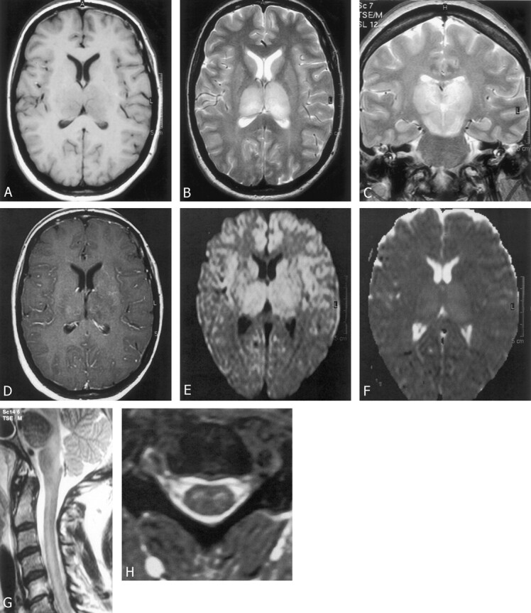
A, Axial spin-echo T1-weighted image (560/14 [TR/TE]) shows hypointensity of the thalami. B, Axial fast spin-echo T2-weighted image (3641/80) showing hyperintensity of the thalami. C, Coronal fast spin-echo T2-weighted image (3641/80) shows hyperintensity of both thalami, red nuclei, and substantia nigra. D, Axial spin-echo T1-weighted image (570/14) reveals subtle heterogeneous enhancement within the thalami. E and F, Single-shot spin-echo echo-planar axial diffusion-weighted image (E) with diffusion sensitivity of b = 1000 s/mm2 (4832/81) demonstrates bilateral thalamic high signal intensity. Corresponding apparent diffusion coefficient map (F) shows increased diffusion in the region of the thalami. This indicates that high signal intensity demonstrated on the diffusion-weighted images represents T2 shine-through rather than cytotoxic edema. G, Sagittal fast spin-echo T2-weighted (2654/100) image of the cervical spine show diffuse hyperintensity of the central cord. H, Axial gradient-echo T2-weighted (697/14; flip angle, 25°) image of cervical spine shows hyperintense gray matter within the cord.
Similar articles
-
Murray valley encephalitis mimicking herpes simplex encephalitis.J Clin Neurosci. 2005 Sep;12(7):822-4. doi: 10.1016/j.jocn.2004.10.010. J Clin Neurosci. 2005. PMID: 16169227
-
Murray Valley encephalitis in an adult traveller complicated by long-term flaccid paralysis: case report and review of the literature.Trans R Soc Trop Med Hyg. 2007 Mar;101(3):284-8. doi: 10.1016/j.trstmh.2006.09.005. Epub 2006 Dec 11. Trans R Soc Trop Med Hyg. 2007. PMID: 17161855
-
Fatal Infection with Murray Valley Encephalitis Virus Imported from Australia to Canada, 2011.Emerg Infect Dis. 2017 Feb;23(2):280-283. doi: 10.3201/eid2302.161161. Emerg Infect Dis. 2017. PMID: 28098530 Free PMC article.
-
The Japanese encephalitis serological group of flaviviruses: a brief introduction to the group.Curr Top Microbiol Immunol. 2002;267:1-10. doi: 10.1007/978-3-642-59403-8_1. Curr Top Microbiol Immunol. 2002. PMID: 12082984 Review. No abstract available.
-
Australian X disease, Murray Valley encephalitis and the French connection.Vet Microbiol. 1995 Sep;46(1-3):79-90. doi: 10.1016/0378-1135(95)00074-k. Vet Microbiol. 1995. PMID: 8545982 Review.
Cited by
-
Transverse myelitis.Neurol Clin. 2013 Feb;31(1):79-138. doi: 10.1016/j.ncl.2012.09.008. Neurol Clin. 2013. PMID: 23186897 Free PMC article. Review.
-
Literature review, case report, and expert discussion of prolonged refractory status epilepticus.Neurocrit Care. 2006;4(1):35-46. doi: 10.1385/NCC:4:1:035. Neurocrit Care. 2006. PMID: 16498194 Review.
-
Brain Imaging in Cases with Positive Serology for Dengue with Neurologic Symptoms: A Clinicoradiologic Correlation.AJNR Am J Neuroradiol. 2018 Apr;39(4):699-703. doi: 10.3174/ajnr.A5544. Epub 2018 Feb 8. AJNR Am J Neuroradiol. 2018. PMID: 29439121 Free PMC article.
-
Clinical and radiological predictors of outcome for Murray Valley encephalitis.Am J Trop Med Hyg. 2013 Mar;88(3):481-9. doi: 10.4269/ajtmh.12-0379. Epub 2013 Jan 7. Am J Trop Med Hyg. 2013. PMID: 23296449 Free PMC article.
-
West Nile virus infection: MR imaging findings in the nervous system.AJNR Am J Neuroradiol. 2005 Feb;26(2):289-97. AJNR Am J Neuroradiol. 2005. PMID: 15709126 Free PMC article.
References
-
- Nash D, Mostashari F, Fine A, Miller J, et al. The outbreak of West Nile virus infection in the New York City area in 1999. N Engl J Med 2001;344:1807–1814 - PubMed
-
- Mackenzie J, Broom AK, Hall RA, et al. Arboviruses in the Australian region, 1990 to 1998. Commun Dis Intell 1998;22:93–100 - PubMed
-
- Kalita J, Misra UK. Comparison of CT and MRI findings in the diagnosis of Japanese encephalitis. J Neurol Sci 2000;174:3–8 - PubMed
-
- Burrow JNC, Whelan PI, Kilburn CJ, et al. Australian encephalitis in the northern territory: clinical and epidemiological features 1987–1996. Aust N Z J Med 1998;28:590–596 - PubMed
Publication types
MeSH terms
LinkOut - more resources
Full Text Sources
Medical