

Case Reports
Peripheral third cranial nerve enhancement in multiple sclerosis
Affiliations
- PMID: 12917135
- PMCID: PMC7973702
Item in Clipboard
Case Reports
Peripheral third cranial nerve enhancement in multiple sclerosis
AJNR Am J Neuroradiol.
2003 Aug.
Abstract
Cranial nerve III dysfunction in multiple sclerosis (MS) is uncommon. Seven cases of isolated cranial nerve III paresis associated with MS have been reported in the English-language literature. MR imaging was obtained in five cases demonstrating lesions within the midbrain. We present the detailed clinical and MR imaging findings of a young woman with MS and an isolated, painful pupil involving complete left cranial nerve III palsy. Initial MR imaging showed isolated enhancement of the cisternal portion of the cranial nerve III, suggesting that peripheral nervous system involvement may develop as part of the disease process in some patients with MS.
Figures
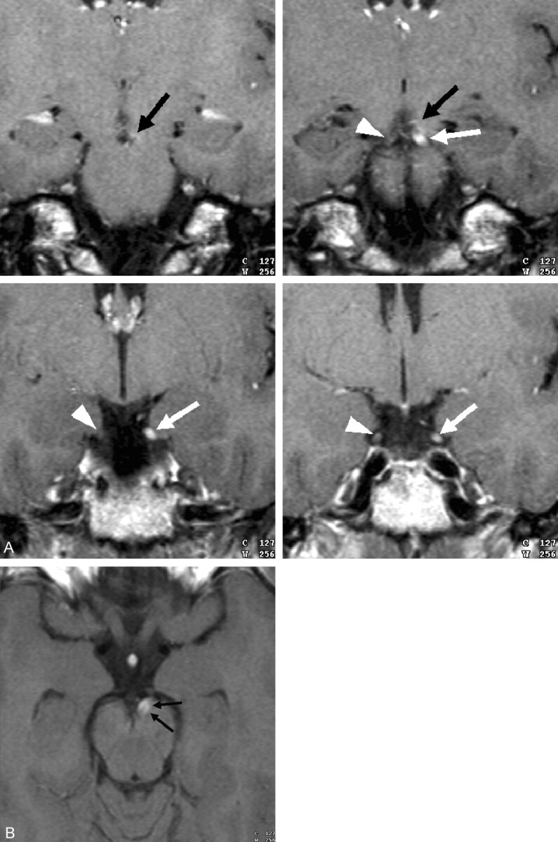
Coronal gadolinium-enhanced T1-weighted images (TR/TE, 600/14) through the cerebral peduncle and cisternal course of cranial nerve III (A) demonstrate marked enhancement and subtle enlargement of cranial nerve III on the left side (white arrows in A). Notice the normal appearance of cranial nerve III on the right (arrowheads in A). There is also subtle enhancement at the cranial nerve III exit zone on the left side (black arrows in A and B), which is better appreciated on the axial gadolinium-enhanced T1-weighted image (B) performed with the same imaging parameters.
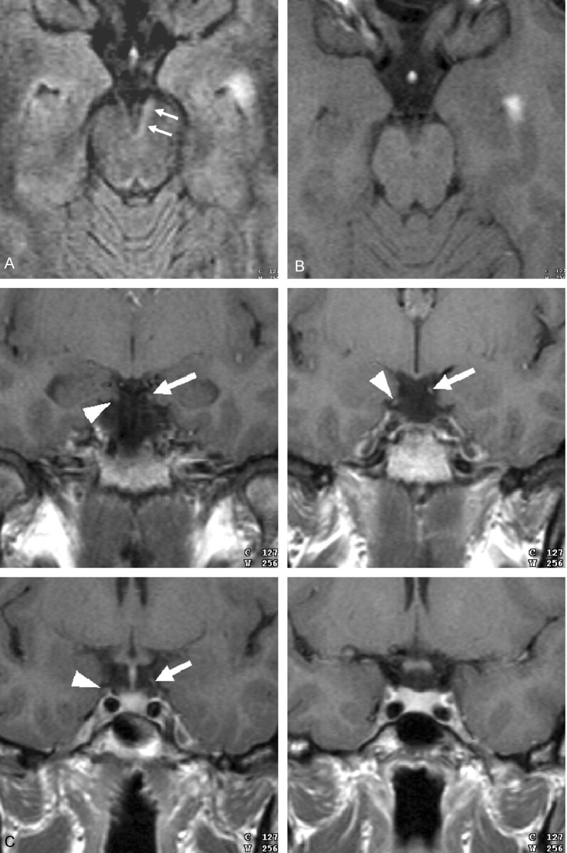
Axial fluid-attenuated inversion recovery (FLAIR) image (TR/TE, 9500/110) through the midbrain (A) demonstrates a subtle area of increased signal intensity (arrows) along the medial margin of the cerebral peduncle on the left side. No enhancement is seen on the axial gadolinium-enhanced T1-weighted image (600/17) (B) in this region. The coronal gadolinium-enhanced T1-weighted images (600/14) through the cisternal course of cranial nerve III (C) demonstrate subtle enhancement of the cranial nerve III on the left side (arrows in C). Notice the normal appearance of cranial nerve III on the right (arrowheads in C).
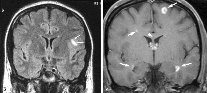
Coronal FLAIR-weighted image (TR/TE, 10,002/168) obtained at the level of the anterior horns of the lateral ventricles shows a focal area of increased signal intensity (arrows) in the subcortical white matter of the inferior frontal lobe on the left side.
Coronal gadolinium-enhanced T1-weighted image (TRTE, 882/14) obtained at the level of the anterior horns of the lateral ventricles shows multiple enhancing white matter lesions (arrows). There is also a nonenhancing lesion in the corpus callosum in midline (arrowhead).
Similar articles
-
Superior divisional third cranial nerve paresis: clinical and anatomical observations of 2 unique cases.Arch Neurol. 2006 May;63(5):771-6. doi: 10.1001/archneur.63.5.771. Arch Neurol. 2006. PMID: 16682550
-
Imaging of the upper cranial nerves I, III-VIII, and the cavernous sinuses.Magn Reson Imaging Clin N Am. 2002 Aug;10(3):415-31, v. doi: 10.1016/s1064-9689(02)00009-0. Magn Reson Imaging Clin N Am. 2002. PMID: 12530227
-
Gd-DTPA enhancement of the cisternal portion of the oculomotor nerve on MR imaging.AJNR Am J Neuroradiol. 1992 Sep-Oct;13(5):1463-70. AJNR Am J Neuroradiol. 1992. PMID: 1414843 Free PMC article.
-
Imaging of Cranial Nerves III, IV, VI in Congenital Cranial Dysinnervation Disorders.Korean J Ophthalmol. 2017 Jun;31(3):183-193. doi: 10.3341/kjo.2017.0024. Epub 2017 May 12. Korean J Ophthalmol. 2017. PMID: 28534340 Free PMC article. Review.
-
Paresis of cranial nerves III, IV, and VI: clinical manifestation and differential diagnosis.Bull Soc Belge Ophtalmol. 1989;237:285-301. Bull Soc Belge Ophtalmol. 1989. PMID: 2486113 Review.
Cited by
-
Magnetic resonance imaging of the cranial nerves in infectious, neoplastic, and demyelinating diseases, as well as other inflammatory diseases: a pictorial essay.Radiol Bras. 2022 Jan-Feb;55(1):38-46. doi: 10.1590/0100-3984.2021.0042. Radiol Bras. 2022. PMID: 35210663 Free PMC article.
-
Cranial Nerve Enhancement in Multiple Sclerosis Is Associated With Younger Age at Onset and More Severe Disease.Front Neurol. 2019 Nov 6;10:1085. doi: 10.3389/fneur.2019.01085. eCollection 2019. Front Neurol. 2019. PMID: 31781014 Free PMC article.
-
Isolated third nerve palsies as the initial manifestation of multiple sclerosis.Neurol Clin Pract. 2018 Oct;8(5):e28-e30. doi: 10.1212/CPJ.0000000000000512. Neurol Clin Pract. 2018. PMID: 30564506 Free PMC article. No abstract available.
References
-
- Kalman B, Lublin FD. Spectrum and classification of inflammatory demyelinating diseases of the central nervous system. Curr Neurol Neurosci Rep 2001;1:249–256 - PubMed
-
- Lucchinetti C, Bruck W, Noseworthy J Multiple sclerosis: recent developments in neuropathology, pathogenesis, magnetic resonance imaging studies and treatment. Curr Opin Neurol 2001;14:259–269 - PubMed
-
- Wingerchuk DM, Lucchinetti CF, Noseworthy JH. Multiple sclerosis: current pathophysiological concepts. Lab Invest 2001;81:263–281 - PubMed
-
- Poser CM. The peripheral nervous system in multiple sclerosis: a review and pathogenetic hypothesis. J Neurol Sci 1987;79:83–90 - PubMed
-
- Waxman SG. Peripheral nerve abnormalities in multiple sclerosis. Muscle Nerve 1993;16:1–5 - PubMed
Publication types
MeSH terms
LinkOut - more resources
Full Text Sources
Medical